
68-year-old lady presented with post-menopausal bleeding for 1 month.
68-year-old lady presented with post-menopausal bleeding for 1 month.
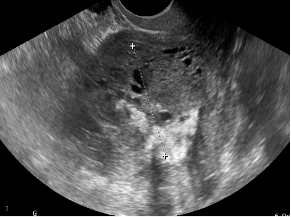

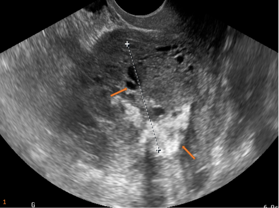
USG PELVIS:
- Grossly thickened endometrium with tiny cystic spaces (Fig. 1).
- Few areas of abnormal vascularity are seen within.
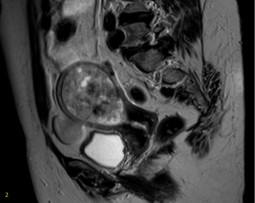
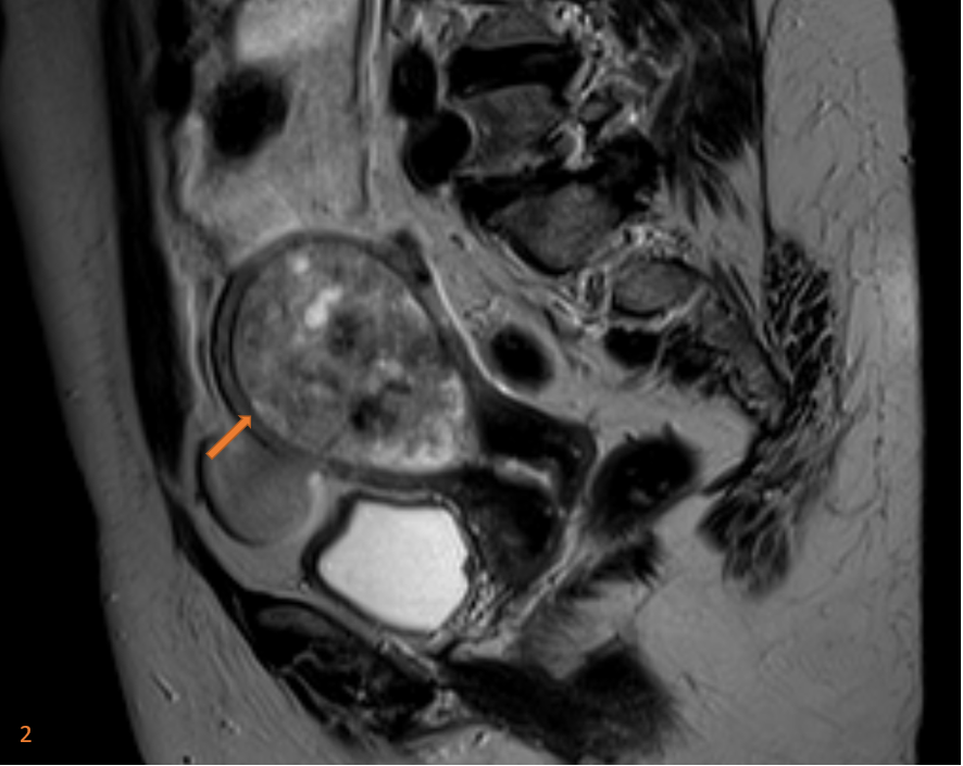
MRI PELVIS:
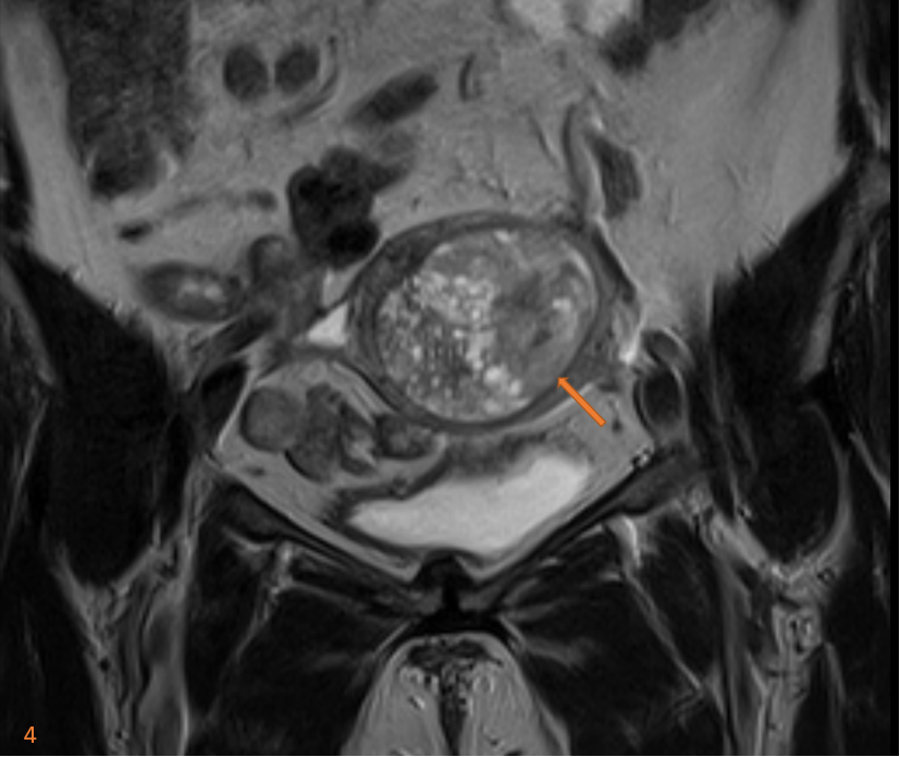
- The endometrium is grossly thickened (thickness measures 4.8 cm) (Fig. 2)
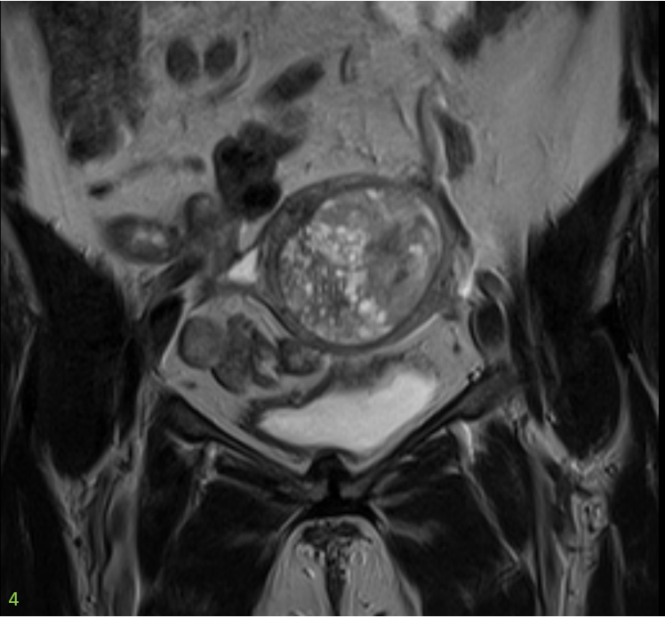
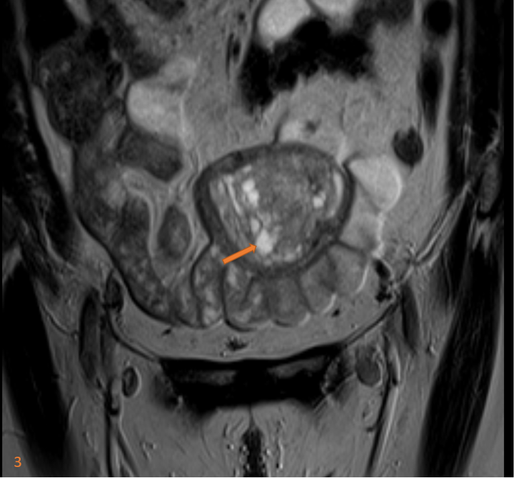
Multiple small T2 hyperintense cystic areas are seen along the right half of the thickened endometrium. (Fig. 3)
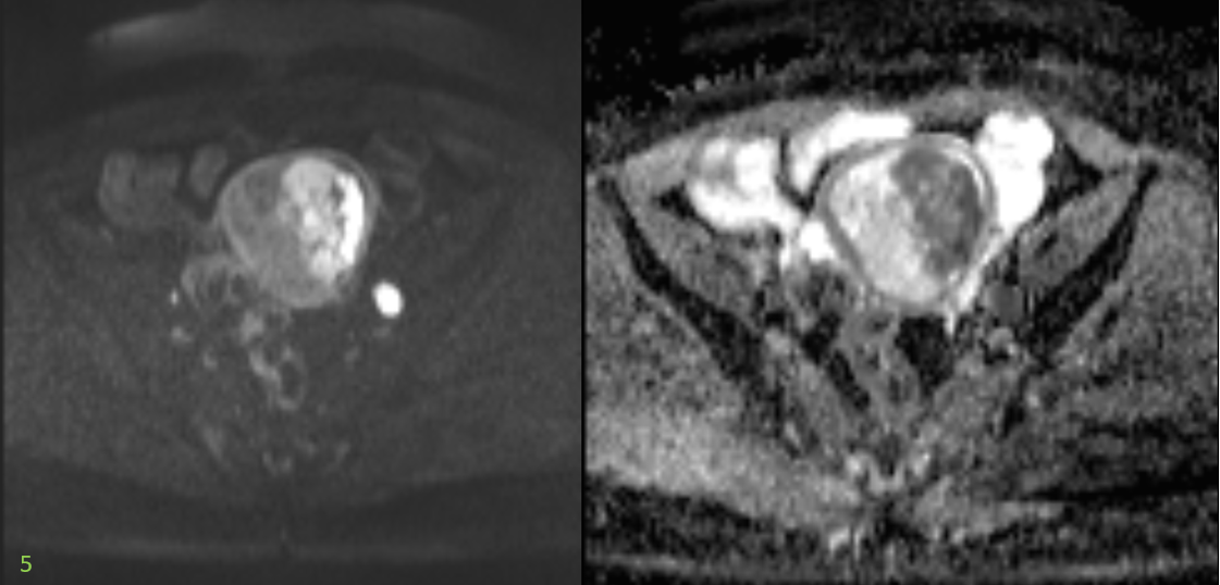
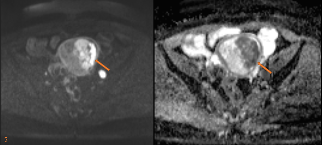
- An ill-defined T1 iso-to hyperintense/ T2 hetero-intense solid appearing lesion with diffusion restriction seen in the left half of the thickened endometrium. (Fig. 4 and 5)
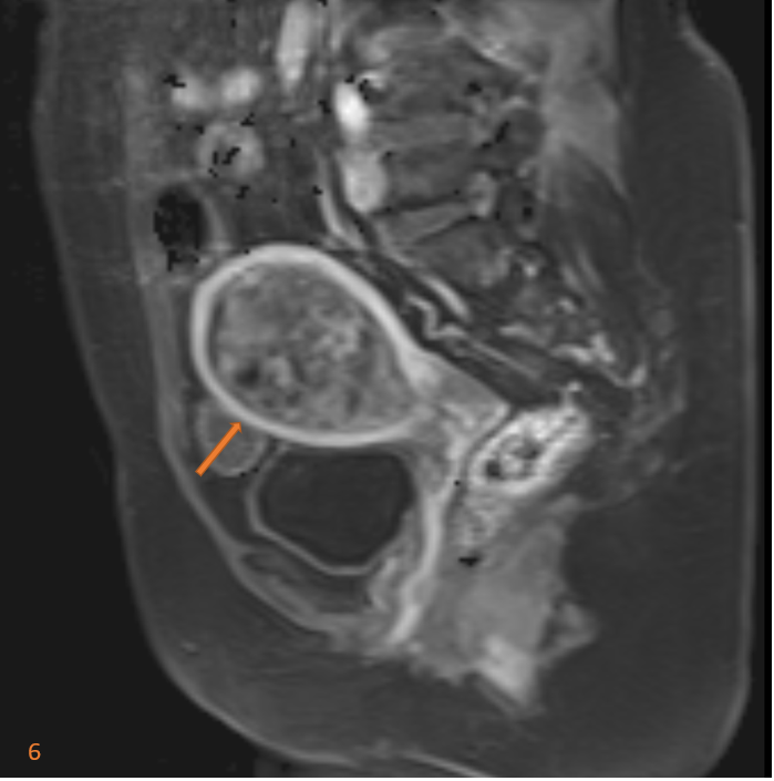
- On dynamic post-contrast images, the lesion shows heterogenous enhancement.
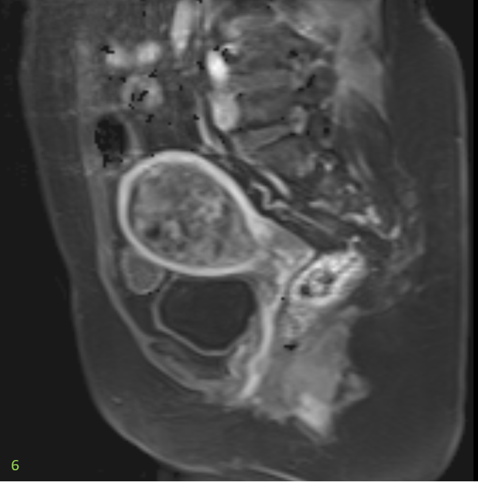
- Myometrium appears intact (Fig. 6)
- Cervical stroma and endocervical canal appear normal.
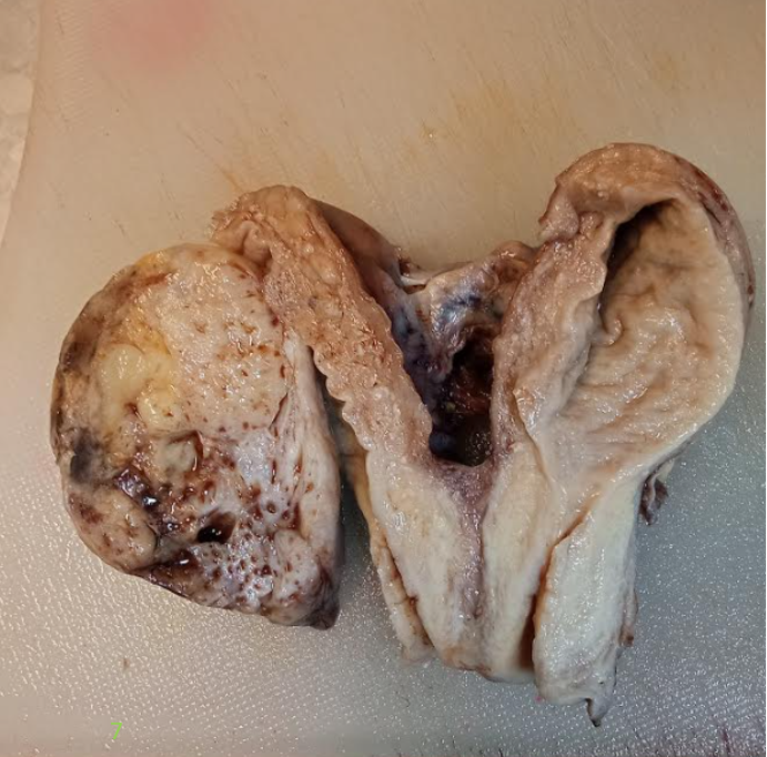
- Endometrial biopsy performed 3 days after the MRI Pelvis demonstrates high-grade malignant tumor (differentials include endometrial carcinoma, serous carcinoma, malignant mixed Mullerian tumor) Gross Hysterectomy specimen with differential tissue components as seen on MRI. (Fig. 7)

DIAGNOSIS:
Malignant mixed Mullerian tumors (MMMT) of the uterus.
DISCUSSION:
- Uterine carcinosarcoma (UCS), also known as malignant mixed Müllerian tumor(MMMT), is a rare mixed epithelial and mesenchymal tumor of the female reproductive tract composed of both high-grade sarcomatous and carcinomatous elements.
- It is comprising only 1-2% of uterine cancers and 3-5% of all uterine malignancies.
- They are the most common variety of mixed epithelial and non-epithelial endometrial tumors, with a clinically aggressive course.
- Three theories proposed to ascertain this tumor’s histogenesis include that MMMTs may be 1) collision tumors, 2) combination tumors, or 3) composition tumors.
- UCS most commonly arises from the uterus but, in rare cases, may originate from the fallopian tube, cervix, or peritoneum.
- UCS is surgically staged in accordance with the 2017 International Federation of Gynecology and Obstetrics (FIGO) Tumor, Node, Metastasis (TNM) classification system.
- Endometrial carcinomas are typically diagnosed at endometrial biopsy or dilatation and curettage.
- MRI is reserved to evaluate the extent of disease.
- MRI is the most accurate imaging technique for the preoperative assessment of endometrial cancer because of its superb soft tissue contrast resolution.
- The routine use of dynamic IV contrast enhancement is necessary for state of-the-art MR evaluation of endometrial carcinoma.

Literature image demonstrating polypoidal mass in the uterine cavity with areas of hemorrhage and necrosis consistent with mixed malignant Mullerian tumor.
REFERENCES:
- Sala E, Wakely S, Senior E et al. MRI of malignant neoplasms of the uterine corpus and cervix. AJR Am J Roentgenol. 2007;188 (6): 1577-87. doi:10.2214/AJR.06.1196 – Pubmed citation
- Teo SY, Babagbemi KT, Peters HE et-al. Primary malignant mixed mullerian tumor of the uterus: findings on sonography, CT, and gadolinium-enhanced MRI. AJR Am J Roentgenol. 2008;191 (1): 278-83. doi:10.2214/AJR.07.3281 – Pubmed citation
- OSR Journal Of Pharmacy (e)-ISSN: 2250-3013, (p)-ISSN: 2319-4219 Www.Iosrphr. Org Volume 3, Issue 9 (October 2013), Pp 49-52
Case contributed by:
Dr. Suvarna Kote
Radiology resident- Columbia Asia Radiology Group.
Dr. Madhu Kumar S B,
Senior Consultant Radiologist
Columbia Asia Radiology Group