
67 year old lady who was diagnosed with breast cancer 5 years ago
A 67-year-old 67-year-old lady who was diagnosed with breast cancer 5 years ago and underwent breast conservative surgery along with adjuvant chemoradiation, has now developed discrete painful lesions in the left upper arm.
Below are the MRI images of the left arm of the patient
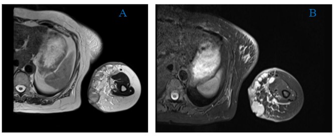
- A- T2 W AXIAL , B- STIR: These demonstrates multiple round to ovoid lesions of varying sizes clustered together which are involving the skin and subcutaneous tissue on the medial aspect of left upper arm. Some of these are demonstrating fluid-fluid levels.
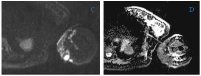
- C-DWI , D- ADC: lesions demonstrates diffusion restriction.
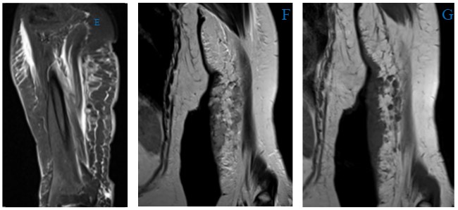
- E- STIR SAG : edematous changes in the left upper limb, suggestive of background lymphadema.
- F-T2 , G- T1 COR: demonstrates few satellite lesions which are hypointense on T1 and hyperintense on T2
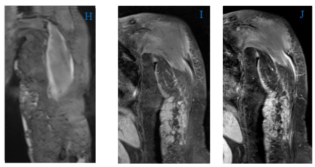
- H- GRE : demonstrates few foci of blooming within the lesions.
- I- T1 pre contrast, J- post contrast sequences: lesion demonstrates moderate heterogenous enhancement.
DIAGNOSIS:
STEWART- TREVES SYNDROME
DISCUSSION:
- Stewart Treves syndrome is a condition where lymphangiosarcoma develops in case of chronic, long-standing lymphedema in the chest wall or upper limb, usually following breast surgeries.
- The reported incidence of developing angiosarcoma in patients surviving at least 5 years post mastectomy and with a history of radiation therapy is 0.07% to 0.45%.
- Exact pathological mechanism is unknown; Stewart and Treves postulated the role of a systemic carcinogenic factor responsible for this process.
- Histologically, the lesion displays networks of small lymphatics and proliferating vascular channels that dissect dermal collagen.
- Clinically they manifest as “spreading bruise,” or a raised purple-red papule, eventually developing tissue infiltration, edema, tumor fungation, ulceration, and even hemorrhage due to increased tumor size.
- DD of cutaneous angiosarcoma: benign and malignant lesions such as hemangioma, hemangioblastoma, squamous cell carcinoma, Kaposi sarcoma, and anaplastic melanoma.
- In our case, a biopsy was done which revealed the underlying pathology to be angiosarcoma.
- Angiosarcoma tends to metastasize by lymphatic or hematogenous pathways, Lungs are the most common site of metastasis.
- Due to the high risk of local recurrence, adjuvant radiotherapy with large doses and wide treatment fields is most often recommended, except in cases where angiosarcomas are radiation-induced.
- The lesion has a poor prognosis, Median survival has been noted at just 7 months.
- Clinical image from literature

- Literature images of HPE of Angiosarcoma

References:
- Murgia RD, Gross GP. Stewart-Treves Syndrome. [Updated 2020 Aug 28]. In: StatPearls [Internet]. Treasure Island (FL): StatPearls Publishing; 2021 Jan-. Available from: https://www.ncbi.nlm.nih.gov/books/NBK507833
- Chopra S, Ors F, Bergin D. MRI of angiosarcoma associated with chronic lymphoedema: Stewart Treves syndrome. Br J Radiol. 2007 Dec;80(960):e310-3. doi: 10.1259/bjr/19441948. PMID: 18065640.
- Smain Nabil Mesli, Amin Khayreddine Ghouali, Fouad Benamara, Fouzi Ahmed Taleb, Hicham Tahraoui, Chakib Abi-Ayad, “Stewart-Treves Syndrome Involving Chronic Lymphedema after Mastectomy of Breast Cancer”, Case Reports in Surgery, vol. 2017, Article ID 4056459, 4 pages, 2017. https://doi.org/10.1155/2017/4056459
HPE images reference:
- Randhawa A, Paul M, Lo S, et al Cutaneous angiosarcoma with lymphoedema: the Stewart-Treves syndrome BMJ Case Reports CP 2019;12:e229101
Dr. Deepti H V, MBBS, DMRD, DNB, EDiR
Consultant Radiologist
Manipal Hospitals Radiology Group.
Dr. Sharath.B.R, MBBS, DMRD, DNB
Radiology Resident
Manipal Hospitals Radiology Group.