
30-year-old lady with complaints of painful swelling over the right foot for past 2-3 months.
- 30-year-old lady with complaints of painful swelling over the right foot for past 2-3 months.
- No history of trauma – fever on and off.
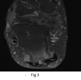
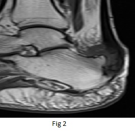
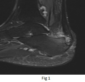
Relatively homogeneous well defined T1 iso/PDFS hyperintense lobulated large irregular shaped lesion involving the navicular bone completely and having destroyed it extends radially in paraosseous, periarticular distribution with well defined punched out areas of lysis/erosions involving cuboid, lateral cuneiform and dorsal/plantar aspects of intermediate cuneiform. It nearly completely encases the head of talus.
DIAGNOSIS:
- GCT of navicular bone.
DISCUSSION:
- Giant cell tumors of bone, also known as osteoclastomas, are relatively common bone tumors and are usually benign. They typically arise from the metaphysis of long bones, extend into the epiphysis adjacent to the joint surface.
- GCT accounts for 5% of all primary bone tumors and 20% of benign skeletal tumors. It peaks during the 3rd decade.
- Most lesions develop in long bones, with the majority of cases occurring about the knee. The bones of the hands and feet are uncommon locations.
- GCT may occur in association with Paget disease, most commonly in the skull, facial bones, pelvis, and spine.
Imaging Features
-best demonstrated on conventional radiographs.
- Shows lytic lesion that has a well-defined but non sclerotic margin, is eccentric in location, extends to the subchondral bone, and occurs in patients with closed physes.
-MRI:
Typical signal characteristics include:
- T1: solid component shows low to intermediate signal,
- T2: heterogeneous high signal with areas of low signal intensity (variable) due to hemosiderin or fibrosis. Fluid-fluid levels can be seen if an aneurysmal bone cyst component is present. High signal in adjacent bone marrow thought to represent inflammatory edema.
- T1 C+: solid components will enhance, helping differentiate from aneurysmal bone cysts. Some enhancement may also be seen in adjacent bone marrow.
- aggressive features: wide zone of transition, cortical thinning, expansile remodeling, or even cortical bone destruction and an associated soft-tissue mass like in our case.
Uncommon Radiologic Manifestations
- Lung metastases have been reported in 1%–6% of cases. Most pulmonary lesions are histologically benign, with an appearance similar to that of the primary bone tumor.
- Rarely, GCT may undergo malignant transformation.
Mimic Lesions
- primary ABC- soft tissue component is absent.
- Plasmacytoma/multiple myeloma.
- Metastasis.
- Chondroblastoma.
- Telangiectatic osteosarcoma and giant cell osteosarcoma.
Dr Naveen SS, MD
Cross sectional imaging fellow MHRG
Dr Dayanand Sagar G, MD
Consultant Radiologist MHRG