
30 year old gentleman presenting with injury to left elbow 2 days back
30-year-old gentleman presenting with injury to left elbow 2 days back.
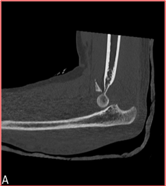

- Figure A demonstrates fracture of coronoid process of ulna with fragment lying antero-superiorly. Posterior dislocation of ulna is also noted.
- Figure B demonstrates mildly displaced fracture of radial head with posterior dislocation of radius at radiocapetallar joint.
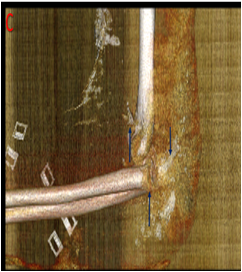
- Figure C ; Volume rendered images clearly demonstrate both radial head and ulnar coronoid process fractures with posterior dislocation at elbow joint.
DIAGNOSIS:
- Complex fracture dislocation at elbow as described above in keeping with Terrible triad of elbow.
Discussion:
- The terrible triad of the elbow is a severe elbow fracture-dislocation pattern and is so-called because it has poor medium-to-long term outcome.
- If inadequately treated; it can result in chronic instability and severe arthritis.
- It occurs predominantly in adults; unlikely to be seen in children.
- Mechanism of injury: Most commonly, a fall onto an outstretched hand, arm in semi-flexion and supination.
- The fall is usually associated with a valgus posterolateral force which disrupts the capsuloligamentous structures sequentially from lateral to medial.

- Computer-generated lateral three-dimensional (3D) view of the elbow demonstrates the normal anatomic configuration of the lateral collateral ligament complex, which includes the LUCL (red), RCL (blue), and annular ligament (yellow).
- Computer-generated medial oblique 3D view shows the normal configuration of the anterior (red), posterior (blue), and transverse (yellow) MCL bundles.
Imaging features
- The terrible triad of the elbow is the association of:
- posterior elbow dislocation
- coronoid process fracture
- radial head fracture
- The posterior elbow dislocation usually involves the ulnohumeral joint.
- The coronoid fracture usually involve the tip or are type I fractures (O’Driscoll Classification).
Points to add in report
- Radial head fracture, coronoid process fracture (however small) with particular attention to anteromedial facet of ulna.
- Radiocapitellar and ulnotrochlear articulation.
- LUCL and MCL tears if possible.
- Status of nerves if assessable.
- Remember that volume rendering and MPR help in accurate estimation of severity of injury.
REFERENCES:
- Mathew PK, Athwal GS, King GJ. Terrible triad injury of the elbow: current concepts. J Am Acad Orthop Surg. 2009;17 (3): 137-51. J Am Acad Orthop Surg (full text) – Pubmed citation.
- Traumatic Elbow Injuries: What the Orthopedic Surgeon Wants to Know Scott E. Sheehan, George S. Dyer, Aaron D. Sodickson, Ketankumar I. Patel, and Bharti Khurana. RadioGraphics 2013 33:3, 869-888
Dr. Ashwini C. MD. FRCR.
Consultant Radiologist
Manipal Hospitals Radiology Group.
Dr. Sushant Mittal. MD
Cross-sectional fellow
Manipal Hospitals Radiology Group.