
" A 26 yearsold gentleman presenting with complaints of knee pain while walking, injury 2 months back due to RTA"
A 26-year-old gentleman presented with complaints of knee pain while walking, injury 2 months back due to RTA.
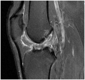
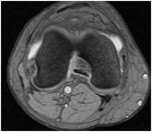
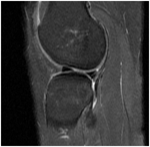
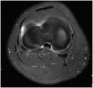
SAGITTAL AND AXIAL PDFS
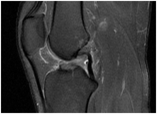
SAGITTAL PDFS
SAGITTAL AND AXIAL PDFS
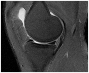
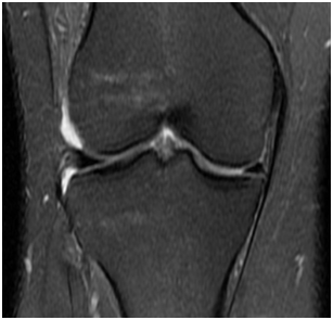
CORONAL PDFS
MRI OF THE RIGHT KNEE JOINT:
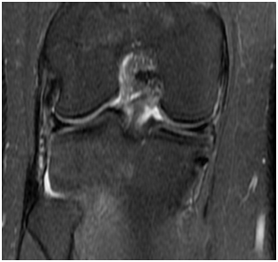
IN THE ABOVE SET OF AXIAL, CORONAL AND SAGITTAL PDFS IMAGES DEMONSTRATES
- High grade partial tear of ACL. Normal posterolateral corner structures ?
- Buckling of Posterior cruciate ligament.
- Anterior tibial translation in relation to femur by 1.1 cm. ?
- Ramp lesion in relation to the posterior horn of medial meniscus ?
- Wrisberg rip tear at the posterior horn of lateral meniscus. ?
- Mild knee joint effusion. Resolving contusion/oedema in the lateral femoral condyle. ?
SAGITTAL AND AXIAL PDFS
SAGITTAL PDFS
SAGITTAL PDFS
SAGITTAL AND AXIAL PDFS
CORONAL PDFS
DIAGNOSIS AND DISCUSSION:
- High grade partial tear of ACL. Anterior tibial translation. Buckling of PCL?
- Ramp lesion in relation to the posterior horn of medial meniscus ?
- Wrisberg rip tear at the posterior horn of lateral meniscus.?
- Minimal knee joint effusion.
RAMP LESIONS:
- Ramp lesions are defined as a vertical(longitudinal) tear of the peripheral capsular attachment of the posterior horn of the medial meniscusat the meniscocapsular junction.
- Most frequently occur in the setting of a pivot shift mechanism of injury (g.anterior cruciate ligament (ACL) injuries) ?
- Complete thin linear fluid signal between the posterior horn of the medial meniscus and posteromedial capsule and posterior meniscal irregularity
WRISBERG RIPS:
- Wrisberg rips, also known as zip tearsor zipper tears, are longitudinal vertical meniscal tears. They occur at the junction of the ligament of Wrisberg and the posterior horn of the lateral meniscus and are commonly associated with anterior cruciate ligament tears . ?
- The ligament of Wrisberg is attached to the lateral aspect of the medial femoral condyle and to the posterior horn of the lateral meniscus, coursing posteriorly to the PCL?
- It is visible on fluid-sensitive sequences, best visualized on sagittal and often on axial images, as a cleft extending anteriorly from the posterior root of the lateral meniscus on several consecutive slices.?
- The pitfall in diagnosing this type of tear is that normally there is a cleft at the attachment site of the ligament of Wrisberg to the posterior horn of the lateral meniscus, which can be mistaken for a tear.
- In our case it is more than 14 mm from the lateral edge of PCL so it should be strongly suspected.
REFERENCES:
- Mohankumar R, White LM, Naraghi A. Pitfalls and pearls in MRI of the knee. AJR Am J Roentgenol. 2014;203 (3): 516-30. doi:10.2214/AJR.14.12969– Pubmed citation
- Xin Liu, Hua Feng, Hui Zhang, Lei Hong, Xue Song Wang, JinZhang. Arthroscopic Prevalence of Ramp Lesion in 868 Patients With Anterior Cruciate Ligament Injury:. (2011) The American Journal of Sports Medicine. 39 (4): 832-7. doi:10.1177/0363546510388933 – Pubmed ?
- Chahla J, Dean CS, Moatshe G, Mitchell JJ, Cram TR, Yacuzzi C, LaPrade RF. Meniscal Ramp Lesions: Anatomy, Incidence, Diagnosis, and Treatment. (2016) Orthopaedic journal of sports medicine. 4 (7): 2325967116657815. doi:10.1177/2325967116657815 – Pubmed ?
- Yujin Yeo, Joong Mo Ahn, Hyorin Kim, Yusuhn Kang, Eugene Lee, Joon Woo Lee, Heung Sik Kang. MR evaluation of the meniscal ramp lesion in patients with anterior cruciate ligament tear. Skeletal Radiology. 47 (12): 1683. doi:10.1007/s00256-018-3007-4
Dr.Rajesh Lavakumar,
MRCS[Ed], FRCR[UK], CCT[UK],
Consultant Radiologist
Manipal Hospitals Radiology Group.
Dr.Ankur Chandra,MD
Cross-sectional fellow
Manipal Hospitals Whitefield