
24 Years, Female presenting with Sudden onset abdominal pain, Sever vomiting
24 years, Female presenting with
- Sudden onset abdominal pain
- Severe vomiting
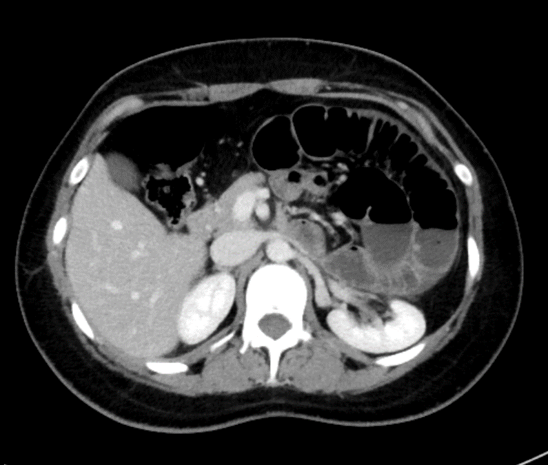
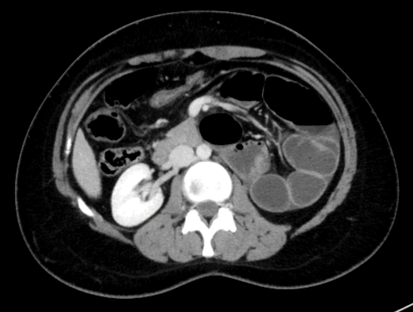
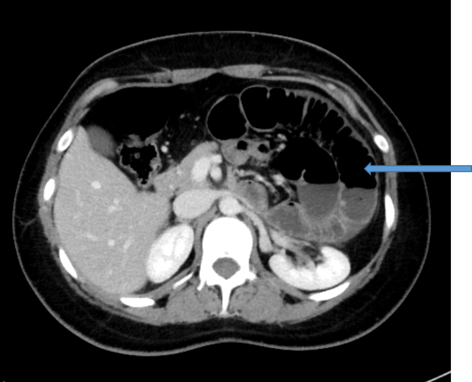
- Post contrast images in axial sections shows dilated jejunal and ileal loops with air fluid levels clustered in left upper abdomen, suggesting small bowel obstruction.
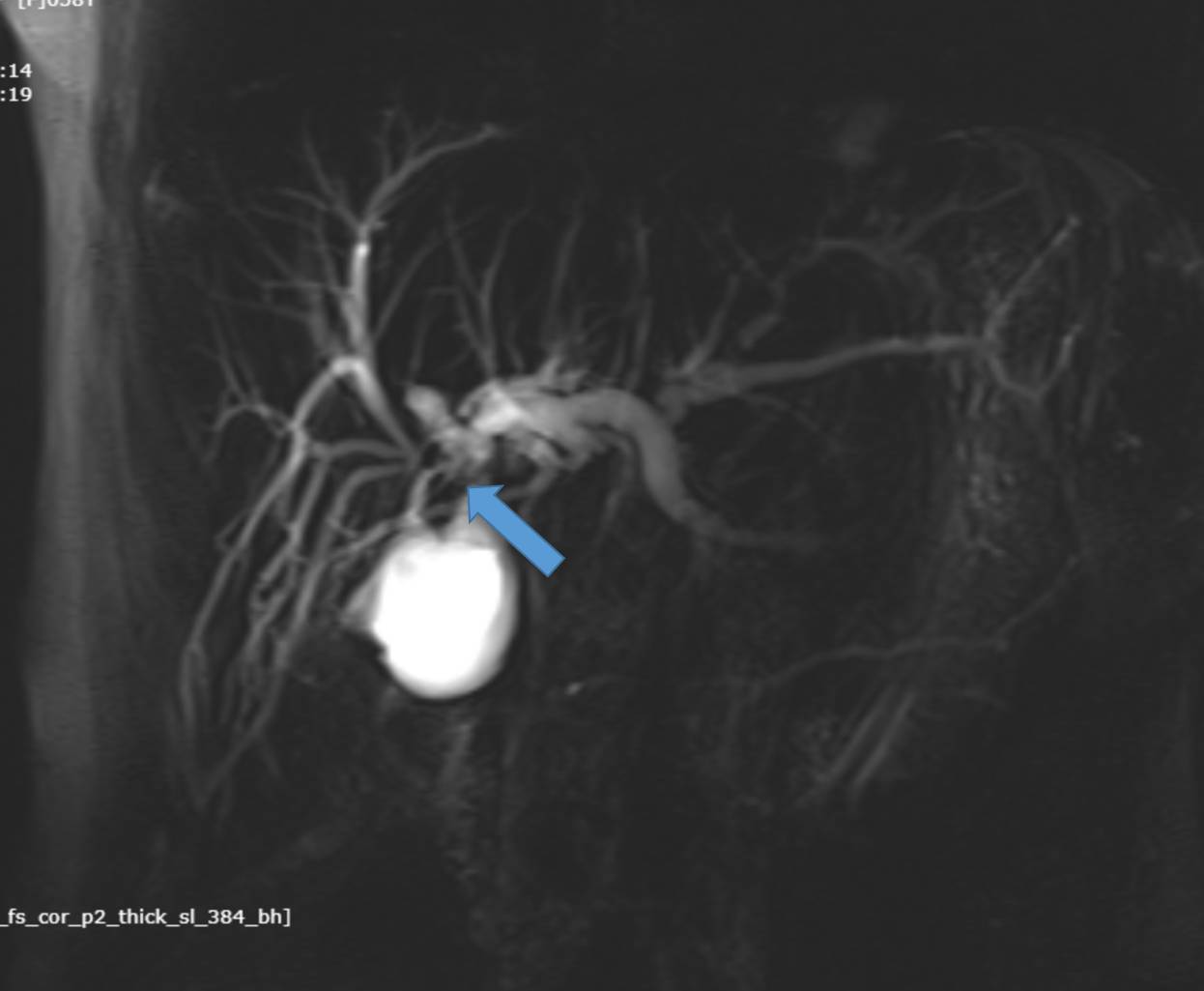
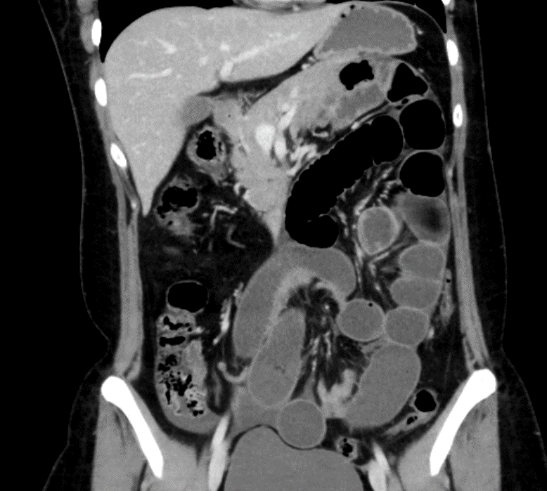
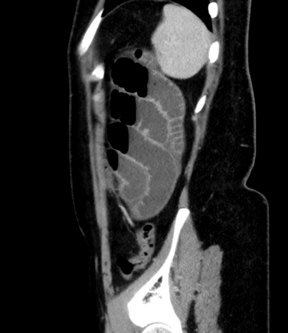
- Post contrast images in coronal and sagittal sections show dilated small bowel loops with air fluid levels and inferior mesenteric vein traversing along bowel loops.
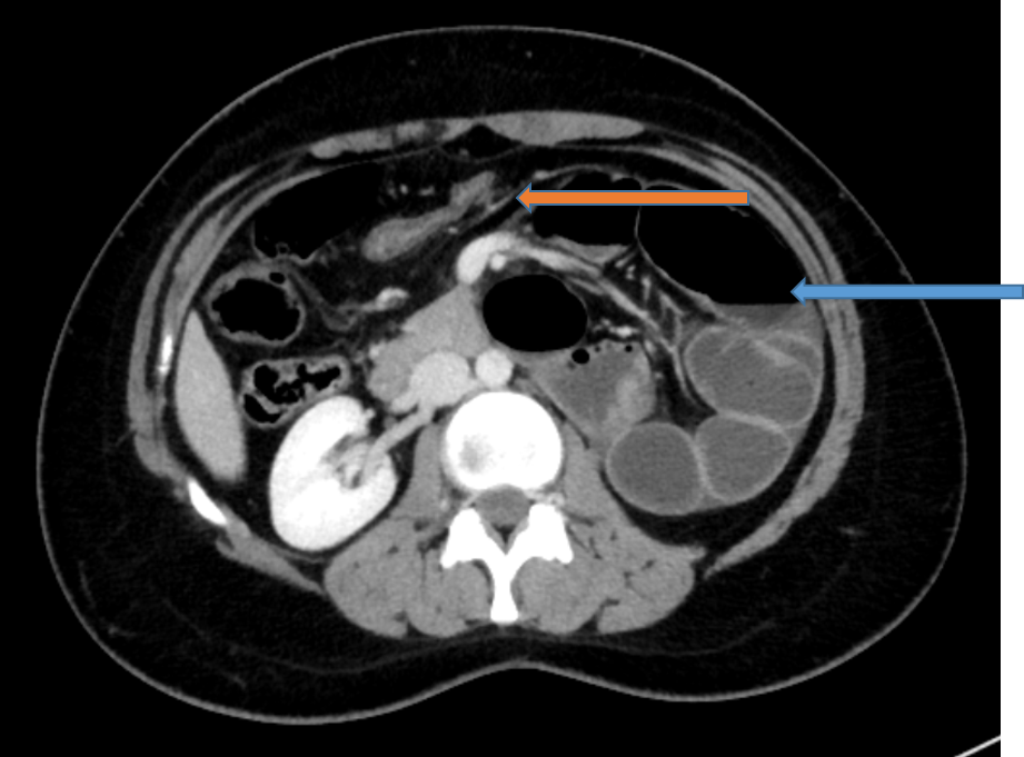
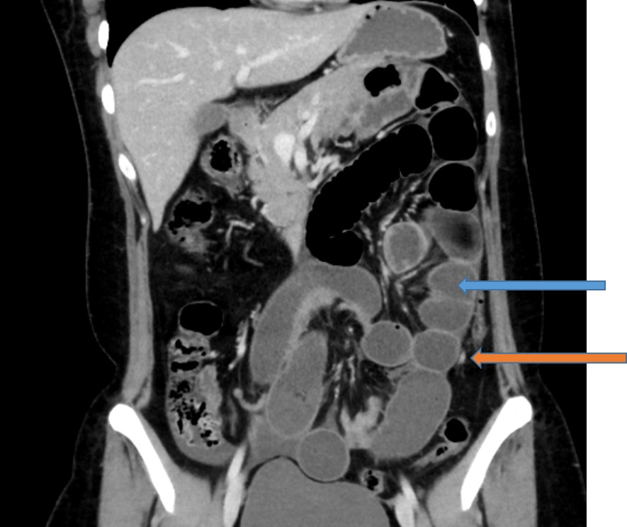
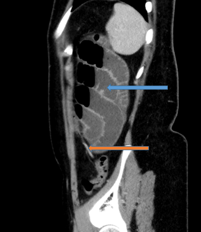
DIAGNOSIS : LEFT PARADUODENAL HERNIA
Discussion:
- Para duodenal hernias, although uncommon, have classically been the most common type of internal hernia.
- These internal hernias may result in closed-loop bowel obstruction.
Pathology:
- They occur when bowel prolapses through Landzert’s fossa, an aperture present in approximately 2% of the population.
- Landzert’s fossa is located behind the ascending or fourth part of the duodenum and is formed by the lifting up of a peritoneal fold by the inferior mesenteric vein and ascending left colic artery as they run along the lateral side of the fossa.
- Small-bowel loops prolapse postero inferiorly through the fossa to the left of the fourth part of the duodenum into the left portion of the transverse mesocolon and descending mesocolon.
Imaging-
RADIOGRAPHS:
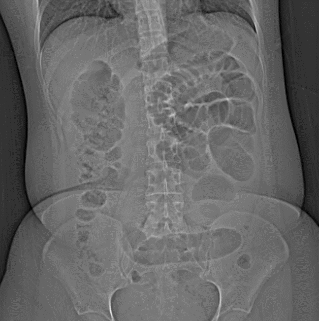
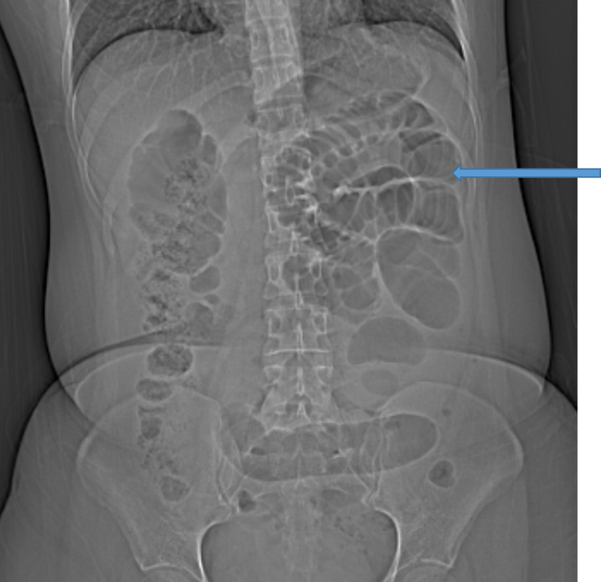
- On radiography or oral contrast studies, these hernias will present as an encapsulated circumscribed mass of a few loops of small bowel (usually jejunal) in the left upper quadrant, lateral to the ascending duodenum.
- These loops may have mass effect, depressing the distal transverse colon and indenting the posterior wall of the stomach.
- Mild duodenal dilatation often occurs, and the efferent loop often shows an abrupt caliber change.
COMPUTED TOMOGRAPHY:
- With CT, similar findings of encapsulated bowel loops are noted, either at the duodenojejunal junction between the stomach and pancreas to the left of the ligament of Treitz; behind the pancreatic tail itself, displacing the inferior mesenteric vein to the left; or between the transverse colon and the left adrenal gland.
- Evidence of small-bowel obstruction with dilated loops and air-fluid levels is also commonly seen.
- There is associated mass effect with displacement of the posterior stomach wall anteriorly, the duodenojejunal junction inferomedially, and the transverse colon inferiorly.
- Mesenteric vessel abnormalities, including enlargement, stretching, and anterior displacement of the main mesenteric trunks, especially the inferior mesenteric vein, to the left, are also helpful findings.
- If the vasculature is optimally visualized, one can often see additional findings of engorged vessels grouped together at the entrance of the hernia sac, with the proximal jejunal arteries showing an abrupt change of direction posteriorly behind the inferior mesenteric artery.
References
- Ghahremani GG. Abdominal and pelvic hernias. In: Gore RM, Levine MS, eds. Textbook of gastrointestinal radiology, 2nd ed. Philadelphia, PA: Saunders, 2000:1993-2009
- Newsom BD, Kukora JS. Congenital and acquired internal hernias: unusual causes of small bowel obstruction. Am J Surg1986; 152:279-284 [Crossref] [Medline]
- Blachar A, Federle MP. Bowel obstruction following liver transplantation: clinical and CT findings in 48 cases with emphasis on internal hernia. Radiology2001; 218:384-388 [Crossref] [Medline]
- Blachar A, Federle MP, Pealer KM, Ikramuddin S, Schauer PR. Gastrointestinal complications of laparoscopic Roux-en-Y gastric bypass surgery: clinical and imaging findings. Radiology2002; 223:625-632 [Crossref] [Medline]
- Meyers MA. Dynamic radiology of the abdomen: normal and pathologic anatomy, 4th ed. New York, NY: Springer-Verlag,1994
- Blachar A, Federle MP. Internal hernia: an increasingly common cause of small bowel obstruction. Semin Ultrasound CT MR2002; 23:174-183 [Crossref] [Medline]
Dr. Srinivas P
Cross-sectional Fellow
Manipal Hospitals Radiology Group (MHRG)
Manipal Hospital, Bengaluru
Dr. Jaishilpa
Senior Consultant Radiologist.
Manipal Hospitals Radiology Group (MHRG)
Manipal Hospital, Bengaluru.