
2 months old presented with complaints of both side facial swelling for 15 days.
History
2 month old presented with complaints of both side facial swelling for 15 days.
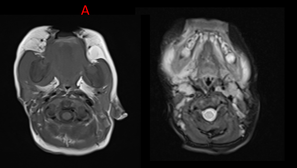
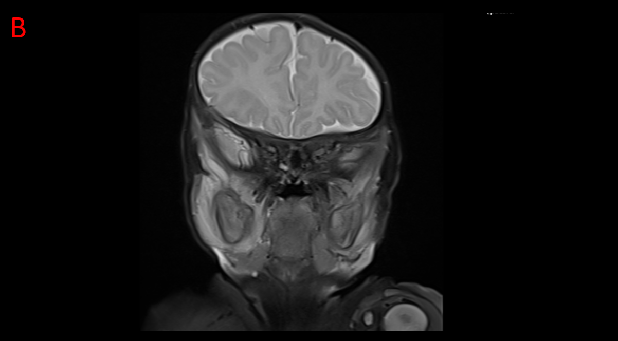
A & B:
T1 ( axial ) and T2 (axial and coronal) shows edema involving bilateral temporalis and masseter muscle along with cortical thickening of the mandible
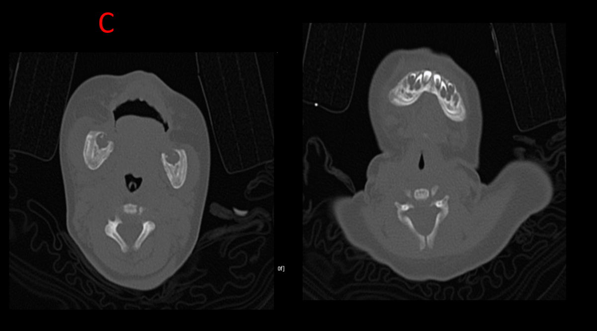
C.
CT shows excessive periostitis with cortical thickening noted involving the mandible
DIAGNOSIS AND DISCUSSION:
Infantile cortical hyperostosis of mandible (Caffey’s disease)
DISCUSSION:
- Caffey disease or infantile cortical hyperostosis is a largely self-limiting disorder which affects infants. It causes bone changes, soft-tissue swelling, and irritability.
- It is distinct from physiological periostitis which can be seen involving the diaphyses of the tibiae, humeri, and femora at the same age.
CLINICAL PRESENTATION:
- Children usually present within the first five months of life with tender and painful soft tissue swelling, erythema, fever, and irritability.
PATHOLOGY:
- Caffey disease is a type I collagenopathy. Both familial and sporadic forms exist.
MARKERS:
- Erthyrocyte sedimentation rate (ESR), C- Reactive protein (CRP) and alkaline phosphatase(ALP) levels are often elevated.
COMMON LOCATION:
The flat bones are most commonly affected:
mandible: in 75-80% of cases
clavicles
scapula: 10% of cases
ribs: lateral aspect
calvaria
Iliac bones
The ulna are the long bones most commonly affected.
Lytic skull lesions have been reported.
The carpals, tarsals, phalanges and vertebral bodies are rarely involved
RADIOGRAPHIC FEATURES:
- Periosteal reaction, either single-layered or lamellated
- subperiosteal cortical hyperostosis
- dense laminated subperiosteal new bone formation
- marked increase in cortical width and density
- in the involved long bones, only the diaphysis is affected, sparing the metaphysis and epiphysis; consequently, the bone becomes spindle-shaped
- soft tissue swelling over the involved bones
MRI usually does not offer much-added value in advancing the diagnosis
Nuclear imaging shows increased radiotracer uptake in the involved bones, both on bone and gallium (Ga-67) scans. The ”bearded infant” appearance refers to intense radiotracer uptake in the mandible
DIFFERENTIAL DIAGNOSIS :
- Osteomyelitis
- Physiological periostitis- Limited to tibia, femur and humerus
- Skeletal dysplasia with osteosclerosis
- Ewing sarcoma
- Metastatic neuroblastoma
- Non – accidental injury
CONCLUSION :
- Caffey disease or infantile cortical hyperostosis is a largely self-limiting disorder which affects infants
- The triad of irritability, swelling, and bone lesions and the presence of mandibular involvement excludes other diagnosis
- Periosteal reaction, , subperiosteal cortical hyperostosis , dense laminated subperiosteal new bone formation are the classical findings
- Common location involved are mandible , clavicles , scapula
REFERENCES :
- Nemec SF, Rimoin DL, Lachman RS. Radiological aspects of prenatal-onset cortical hyperostosis [Caffey Dysplasia]. European journal of radiology. 81 (4): e565-72. doi:10.1016/j.ejrad.2011.06.049– Pubmed
- Glorieux FH. Caffey disease: an unlikely collagenopathy. The Journal of Clinical Investigation. 115 (5): 1142-4. doi:10.1172/JCI25148– Pubmed
- Nistala H, Mäkitie O, Jüppner H. Caffey disease: new perspectives on old questions. Bone. 60: 246-51. doi:10.1016/j.bone.2013.12.030– Pubmed
- Lachman RS. Taybi and Lachman’s Radiology of Syndromes, Metabolic Disorders, and Skeletal Dysplasias. ISBN: 9780323019316
- Sanders DG, Weijers RE. MRI findings in Caffey’s disease. Pediatric radiology. 24 (5): 325-7. Pubmed
- Bykov S, Garty I, Spiegel R, Lumelsky D, Horovitz Y. “Bearded infant” appearance on bone and Ga-67 scintigraphy in a child with localized mandibular Caffey’s disease. Clinical nuclear medicine. 28 (5): 426-8. doi:10.1097/01.RLU.0000063862.89622.BF– Pubmed
- . Brand RA. Biographical sketch: John Caffey, MD (1895-1978). (2011) Clinical orthopedics and related research. 469 (3): 753-4. doi:10.1007/s11999-010-1665-1– Pubmed
Dr. SRIRAM PATWARI
Senior Consultant Radiologist
Manipal Hospital, Yeshwanthpur, Bengaluru.
Dr. SHARNITHA JOHNSON
Senior resident
Manipal Hospital, Yeshwanthpur, Bengaluru.