
2-day old infant, a DCDA twin, presented with respiratory distress syndrome. Antenatal scan showed a large splenic lesion since early pregnancy, with interval scans showing gradual progressive in size. Mother is an elderly primipara, with previous history
HISTORY
- 2-day old infant, a DCDA twin, presented with respiratory distress syndrome. The antenatal scan showed a large splenic lesion since early pregnancy, with interval scans showing gradual progress in size. Mother is an elderly primipara, with a previous history of hypothyroidism and cervical cerclage.
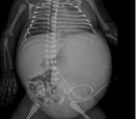
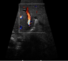
A. On AP abdomen there is distension of abdomen on the left with displacement of bowel loops to right side – likely due to mass lesion.On USG, there was a large heterogeneously hypoechoic mass occupying the left hemi-abdomen indenting on the liver, spleen and displacing the small bowel loops to the right lower quadrant.
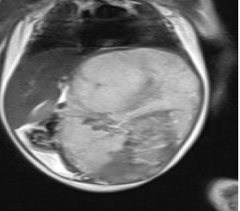
B. T2 and T1 demonstrate a well-defined T2 heterointense abdomino-pelvic mass lesion occupying the left hemi-abdomen, arising from left lobe of liver.
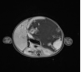
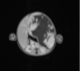
C. T1 post-contrast images demonstrate progressive centripetal contrast enhancement with gradual filling in.
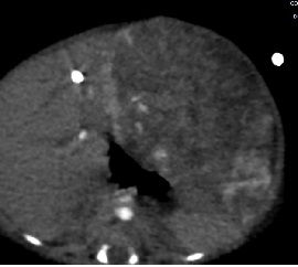
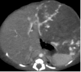
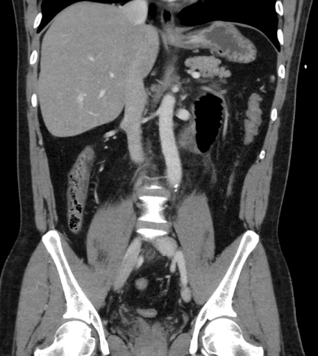
D. CECT abdomen confirms the origin of the lesion from left lobe of liver. Spleen is visualized separately and is normal.
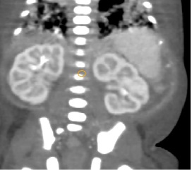
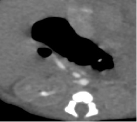
E. CECT abdomen demonstrates moderate narrowing of the infrarenal aorta with multiple collaterals from the left hepatic artery, and branches of left external and internal iliac arteries.
DIAGNOSIS:
- INFANTILE HEMANGIOENDOTHELIOMA
DISCUSSION:
- Infantile hemangioendothelioma is the most common liver tumor during the first 6 months of life.
- On CT, infantile hemangioendothelioma is a well-defined mass that is hypoattenuating relative to the normal liver parenchyma with progressive centripetal contrast enhancement. In larger tumors, central enhancement is often lacking due to fibrosis, hemorrhage, or necrosis.
- A dilated and elongated hepatic artery and early filling of dilated draining hepatic veins may be seen with significant arteriovenous shunting. Because
- Due to increased vascular supply to these tumors, a striking decrease in the aortic caliber is often seen distal to the celiac artery origin.
- Most important alternative diagnosis to infantile hemangioendothelioma in this age group is hepatoblastoma.
- Hepatoblastoma demonstrates irregular hypoattenuation and calcifications, appearing less fine and granular than haemangioendothelioma. CECT typically demonstrates inhomogeneous intratumoral uptake that is generally less intense than in the normal liver parenchyma and hemangioendothelioma.
- Other differentials include mesenchymal hamartoma and metastatic neuroblastoma.


Left external iliac angiogram shows feeders from the left circumflex artery to the lesion which was embolized
REFERENCES
- Roos JE, Pfiffner R, Stallmach T, Stuckmann G, Marincek B, Willi U. Infantile hemangioendothelioma. Radiographics. 2003 Nov;23(6):1649-55.
Dr Rajesh V Helavar
Senior Consultant Radiologist
Manipal Hospital, Yeshwanthpur, Bengaluru
Dr Akshay K
Radiology resident
Manipal Hospital, Yeshwanthpur, Bengaluru
Dr Madhu Kumar
Senior Consultant Radiologist
Manipal Hospital, Yeshwanthpur, Bengaluru
Dr Vikhyath Shetty
Consultant radiologist
Manipal Hospital, Yeshwanthpur, Bengaluru