18 year old male with right shoulder pain for 1 month Few episodes of fever for 1 week
18-year-old male with right shoulder pain for 1 month Few episodes of fever for 1 week
FINDINGS
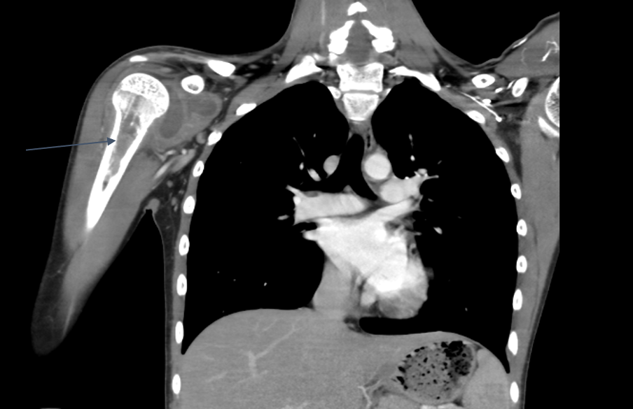
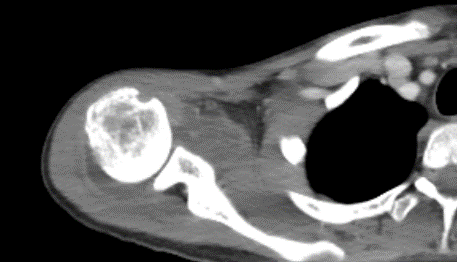
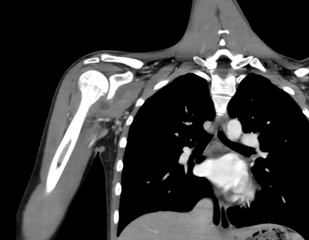
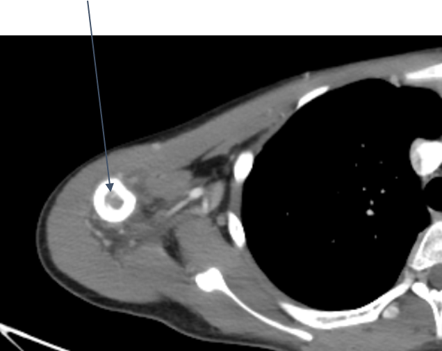
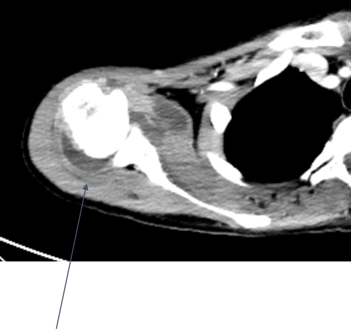
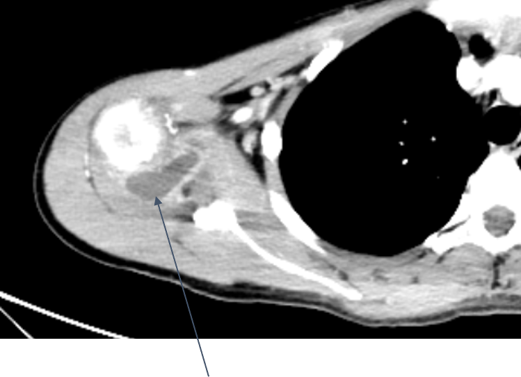
- Multiple patchy mixed lytic-sclerotic areas are seen within the marrow cavity of the meta-diaphyseal region of the proximal right-sided humerus.
Multiple skip lesions were noted in the shaft of the right humerus. - Small peripherally enhancing hypodense well-encapsulated fluid collection seen in right shoulder joint suggestive of synovitis.
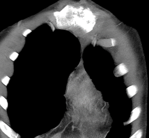
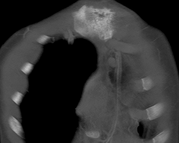
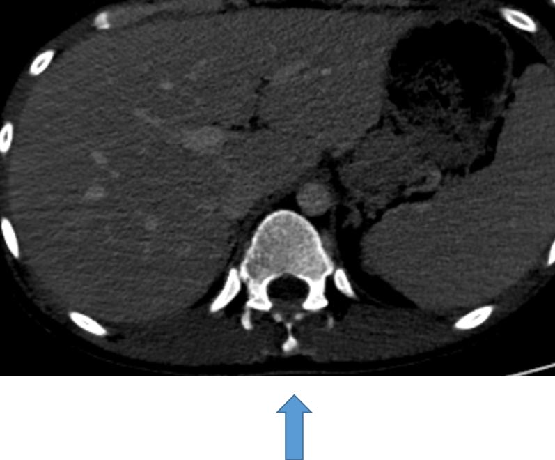
- Area of mixed lytic-sclerotic lesions seen in manubrium sternum on the left side with associated soft tissue mass.
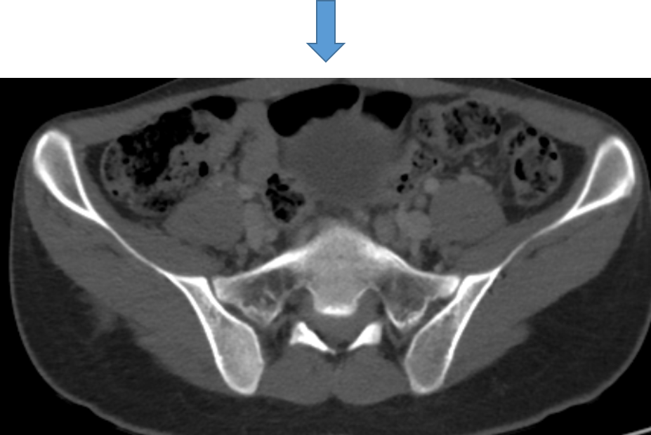
- Similar bone changes with associated epidural soft tissue were seen involving the D12 spinous process and S1 segment.
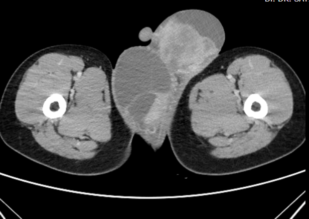
- Diffusely enlarged bilateral testes with surrounding multiloculated hydrocele.
Both testes are replaced by heterogeneously enhancing mass lesions
DIAGNOSIS:
- Diffusely enlarged testes with surrounding multiloculated hydrocele, with testicles being replaced by heterogeneously enhancing mass lesions. Multifocal bone lesions involving right proximal humerus, left manubrium sterni, D12 vertebra and S1 segment with associated soft tissue masses as described. Ultrasound revealed enlarged heterogeneous echotexture of testicles with multiple hypoechoic areas within and increased vascularity.
- Multiple enlarged lymph nodes noted in the para-aortic, retroperitoneal and superficial inguinal regions.
Appearances suggest a haematological malignancy.
FINAL DIAGNOSIS:
Acute myeloid leukemia (monocytic variant) with multiple myeloid sarcomas (Bone marrow biopsy)
DISCUSSION:
- Chloromas usually occurs as a secondary manifestation, either before or simultaneously with AML. Less frequently, they may appear after complete hematologic remission, which strongly indicates bone marrow or other extramedullary relapse.
- The development of a granulocytic sarcoma in the setting of a chronic myeloproliferative disorder or a myelodysplastic syndrome signifies a blast crisis and is associated with disease progression and a poor prognosis.
- On gray-scale ultrasonography, chloromas appear hypoechoic and relatively homogeneous.
- Chloromas may have “septations”, which appear as hypoechoic lines radially extending inside the tumor. Testicular seminomas may also present with septations, usually hyperechoic.
- Bony lesions are more common in leukemic cases. One of the most important radiological findings is metaphyseal lucent band.
- Osteosclerotic lesions, which are considered as bone marrow infarct or may be due to leukemic cell infiltration in the bone marrow may be seen.
- The skull has been an uncommon site of involvement; however, periosteal reaction and osteolytic lesions have been reported.
COMMON MUSCULOSKELETAL FINDINGS:
- Lucent Metaphyseal Bands
- Periosteal Reaction
- Leukemic Infiltration of Bone Marrow
- Pathologic Fracture
- Primary bone lymphoma
- Granulocytic sarcoma
- Compartment Syndrome
REFERENCES
- Bony Lesions in Pediatric Acute Leukemia: Pictorial Essay Makhtoom Shahnazi et al.
- Imaging Musculoskeletal Manifestations of Pediatric Hematologic Malignancies – American Journal of Roentgenology.
- The Bone Changes of Leukemia in Children – Radiographics.
Dr.PRIYANKA ROUT
Radiology resident
Manipal Hospital, Yeshwanthpur, Bengaluru.
Dr. M MATHEW JAMES
Radiology resident
Manipal Hospital, Yeshwanthpur, Bengaluru.