
18 year old male with history of right sided facial pain
18-year-old male with a history of right-sided facial pain
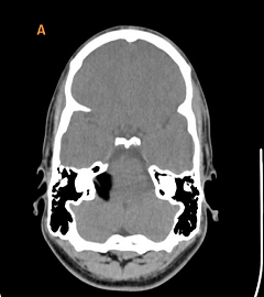


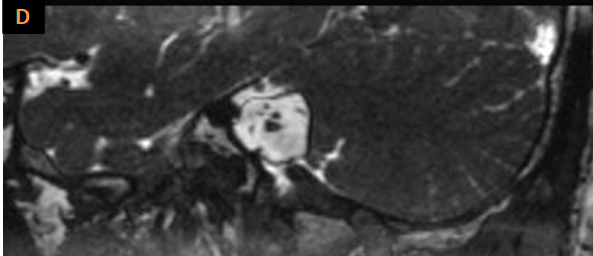
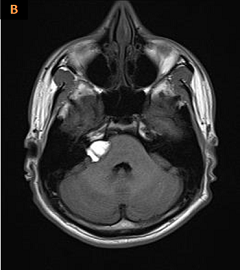
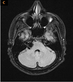
CT image (A) shows a well-defined extra-axial hypodense lesion with fat density in the right cerebellopontine angle. The lesion is hyperintense on T1 (B) with suppression on FLAIR (C). There is the encasement of the VII- VIIIth nerve complex (arrow) which is well demonstrated on sagittal CISS image (D)


DIAGNOSIS:
Cerebellopontine angle Lipoma
DISCUSSION:
- Lipomas are uncommon differential for Cerebellopontine angle (CPA) masses, which are thought to originate from persistence and mal-differentiation of the meninx primitiva.
- Coexisting intravestibular lipomas are known to occur with CPA lipomas; however, there is no reported association with other brain malformations.
- Lipomas appear as homogeneously hypoattenuating lesion with a negative attenuation value on CT and as characteristic and suggestive homogeneous high signal intensity on T1w images, with complete suppression on fat-suppressed images and absence of post contrast enhancement. Therefore, nonenhanced T1-weighted imaging should be performed when evaluating a CPA syndrome to look for spontaneous hyperintense lesions, such as lipomas.
- Unlike vestibular schwannomas, surgical resection of CPA lipomas is difficult to achieve and not frequently indicated. These tumors are indolent, but infiltrate along cranial nerves, making complete removal difficult due to the high risk of postoperative cranial nerves deficit.
Dr. Sriram Patwari
MD, PDCC (Neuroradiology)
Consultant Radiology, Co-lead Neuroradiology
Manipal Hospitals Radiology Group