A 65 year old male, Presents with low back ache with bilateral lower limb pain.
- TRASTFINDINGS – MRI LUMBAR SPINE WITHOUT IV CON
- FINDINGS – MRI LUMBAR SPINE WITHOUT IV CONTRAST
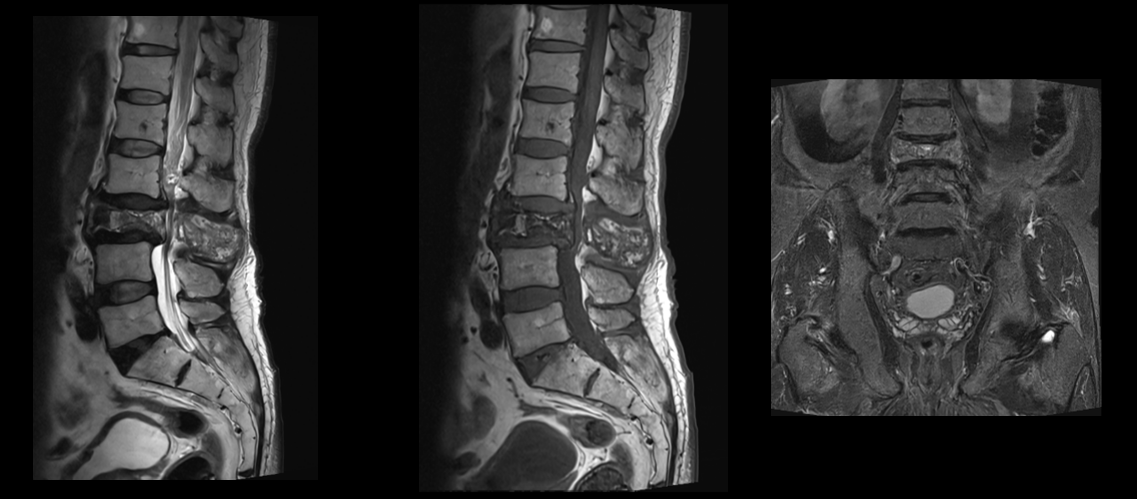
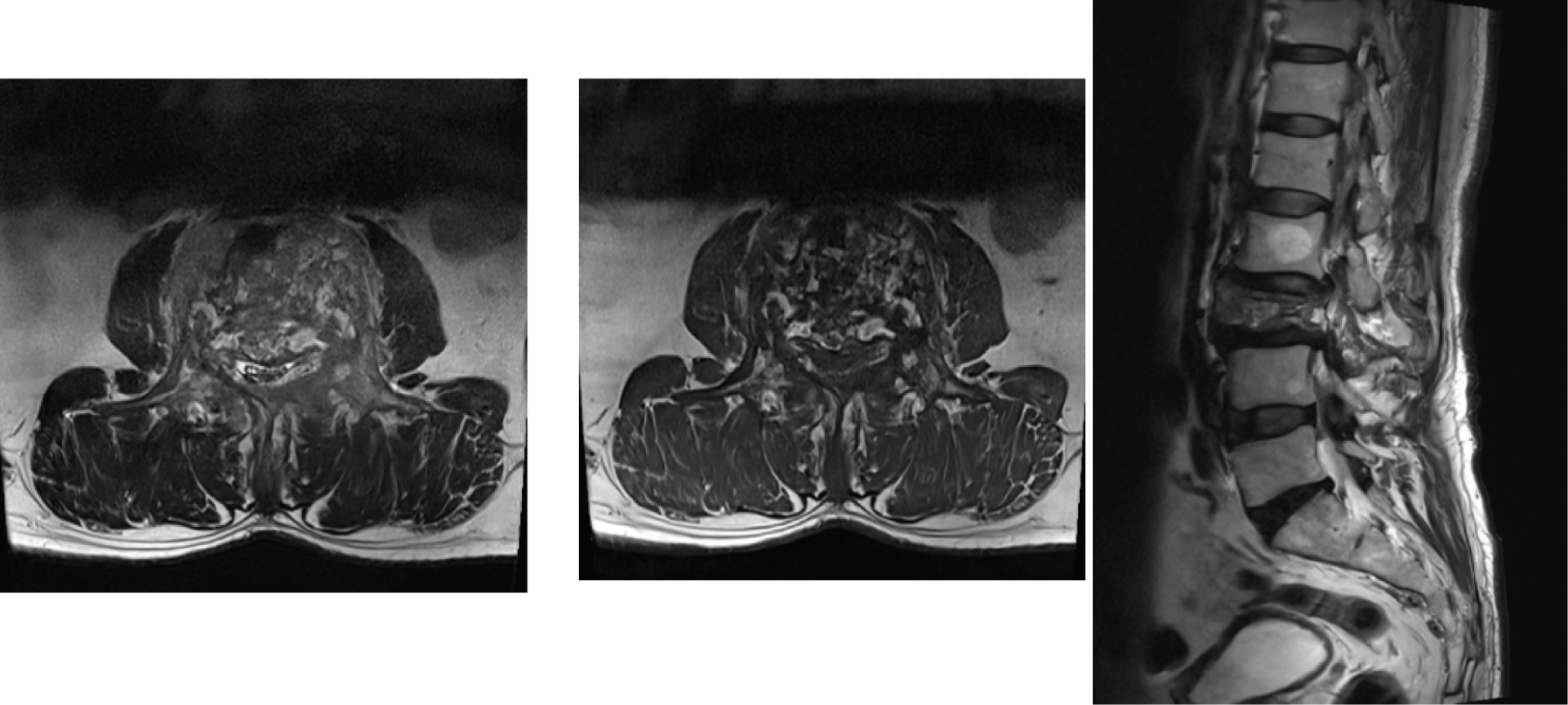
Mixed signal intensity lesion with interspersed fat signal intensity areas is seen involving L3 vertebral body and the posterior elements causing bony expansion. There is redundancy of the cauda equina nerve roots is seen proximally. There is moderate reduction in the height of L3 vertebral body of up to 30%. No aggressive soft tissue is seen in the surrounding region. No inflammatory changes soft tissue oedema is seen in the adjoining paravertebral regions. No collections.
There is resultant severe spinal canal narrowing and compression of the thecal sac/cauda equina nerve roots. Incidental benign vertebral body haemangioma is seen at L2 level.
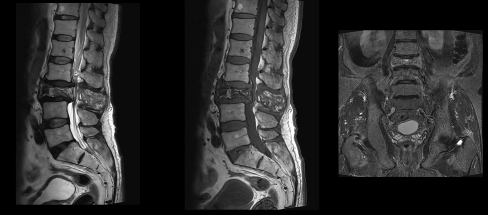
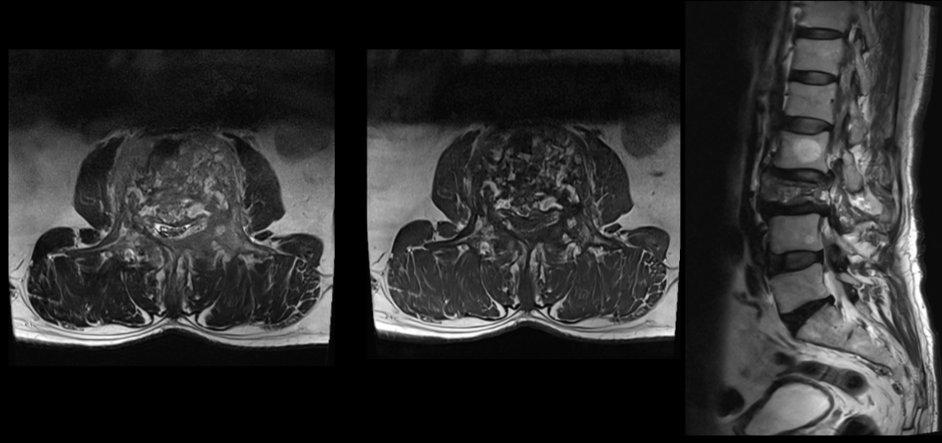
Paget’s disease of the vertebral body.
DISCUSSION:
Paget’s disease (PD) or osteitis deformans is a chronic metabolically active bone disease, characterized by a disturbance in bone modelling and remodelling due to an increase in osteoblastic and osteoclastic activity.
The vertebra is the second most commonly affected site (first being the pelvis). In PD the loss of homeostatic control leads to increased osteoblastic and osteoclastic activity and constitutes the background for the main three phases:
- The initial lytic phase - mainly osteoclastic activity
- The late osteoblastic phase - new bone formation
- The intervening mixed phase - combination of osteoblastic and osteoclastic activities.
- One more phase - “inactive sclerotic phase”- characterised by normal or decreased bone activity, has also been described when the stimulation of new osteoblast and osteoclast formation ceases.
These phases can be evident in the same patient and at the same time in different bones including the vertebral column.
The pathomechanisms and the dynamics involved in bone remodelling in PD
The enhanced abnormal osteoblastic activity results in periosteal and endosteal new bone formation (apposition).
The abnormal osteoclastic activity on the endosteal surface results in bone resorption (absorption). The various combinations of these mechanisms give rise to four different patterns of bone remodelling at the periosteum/endosteal interface leading to bone enlargement:
- Periosteal and endosteal apposition- the apposition on the periosteal side results in vertebral body enlargement, but the bone marrow space is decreased
- Periosteal apposition and endosteal absorption- new bone formation predominates on the periosteal surface and it is responsible for the vertebral body enlargement, while the absorption on the endosteum results in an increased bone marrow space
- Periosteal apposition with normal endosteal surface- the apposition on the periosteal side results in vertebral body enlargement, but the bone marrow space is normal in size
- Focal periosteal apposition –“pumice stone” appearance
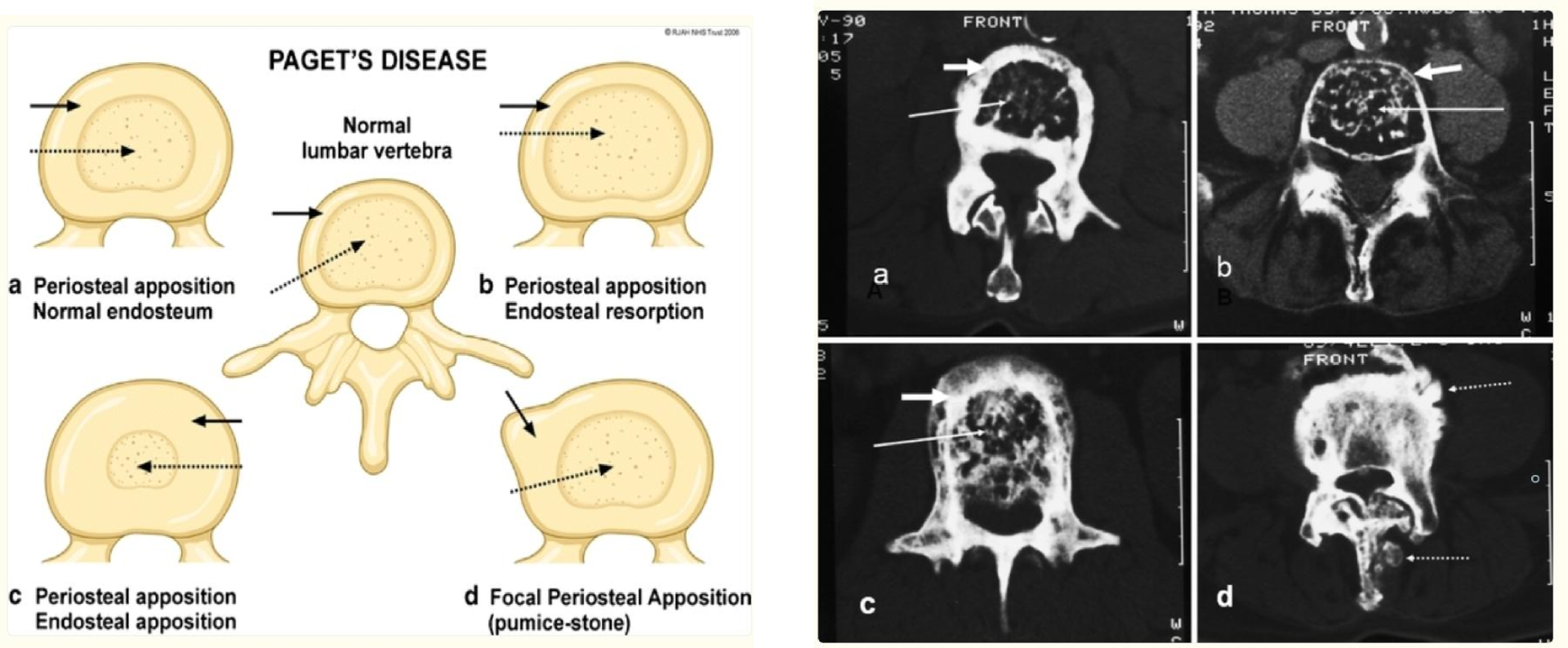
The osseous mechanisms involved in vertebral body enlargement in Paget’s disease and its effect on the size of the marrow (dashed arrows) and cortex (solid arrows).
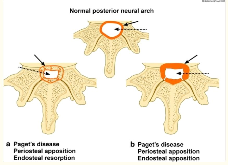
Diagram showing the periosteal and endosteal Pagetic osseous mechanisms involving the cortex of the spinal canal resulting in spinal canal narrowing.
Normal cortical thickness (orange) of the spinal canal (white) is depicted at the top.
a - Expansion of bone due to periosteal apposition/endosteal resorption results in a thin cortical outline (solid black arrow) of the narrowed spinal canal (dashed arrow).
b - Bony expansion due to periosteal apposition/endosteal apposition results in a thickened cortical outline (solid black arrow) of the narrowed spinal canal (dashed arrow)
Osseous changes in vertebral body
The radiological appearance of vertebral body expansion is characterised on radiographs by an increase in the anteroposterior and lateral vertebral dimensions. However, the height of the vertebra is unchanged.
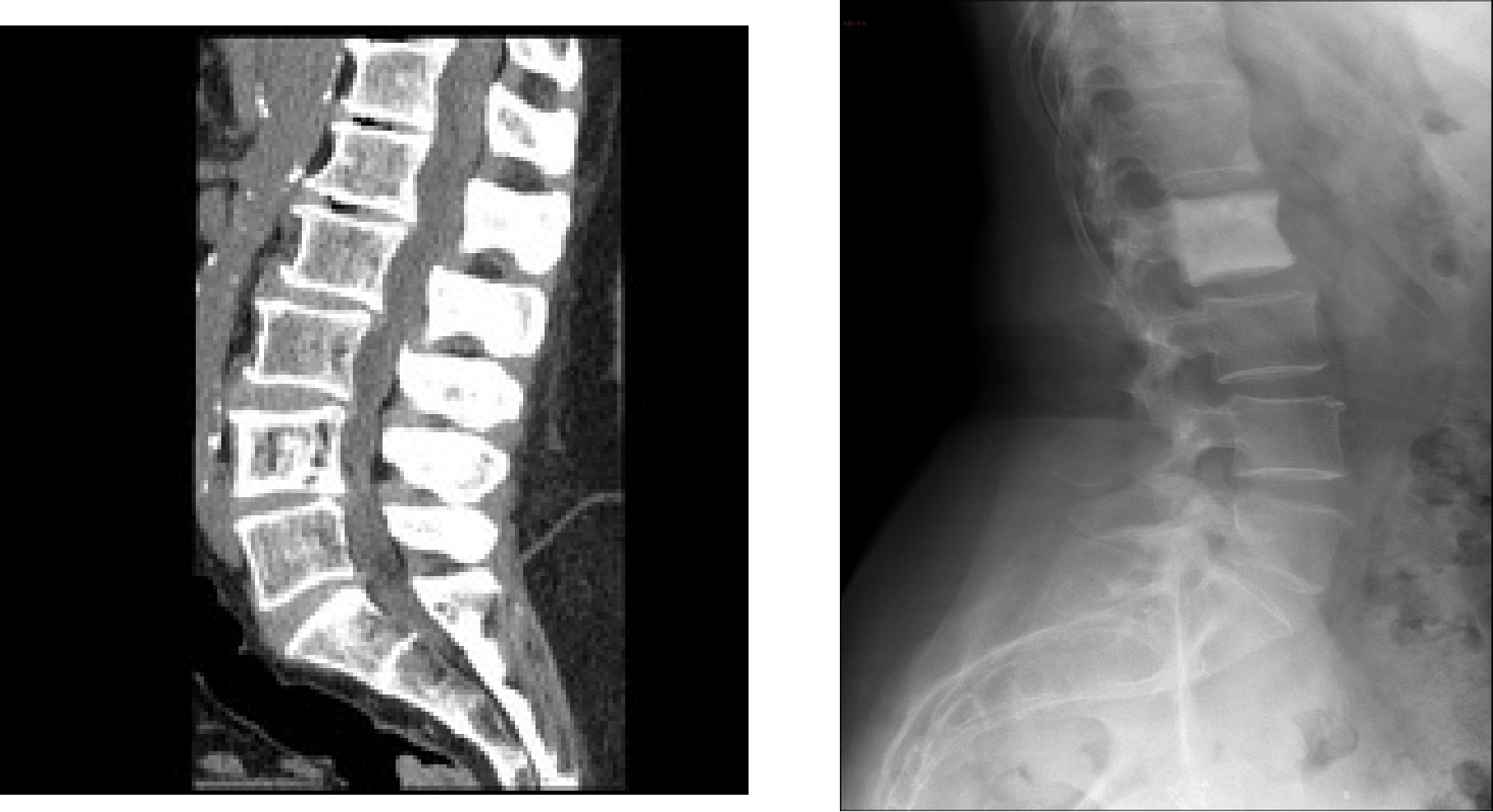
- The combination of trabecular bone hypertrophy and thickening at the end-plates with apposition/absorption on the periosteal/endosteal surfaces at the anterior and posterior vertebral borders leads to the “picture frame” sign.
- Progression of the sclerotic phase in the spine leads to “ivory vertebra”, due to an increase in the density of the vertebral body, denser than the normal vertebral bodies.
CT
CT conspicuously exhibits the classic findings of Paget disease that include osteolysis, trabecular coarsening, cortical thickening, and osseous expansion.
CT is helpful in the workup of suspected complications including fractures, spinal stenosis, and secondary neoplasms.
MRI
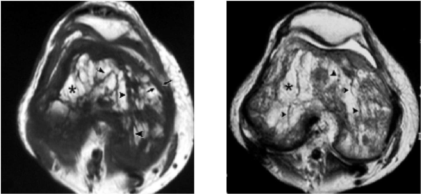
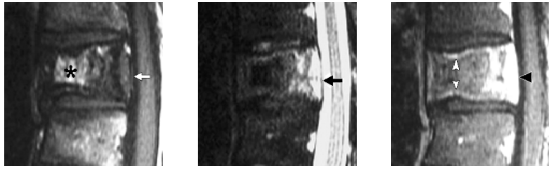
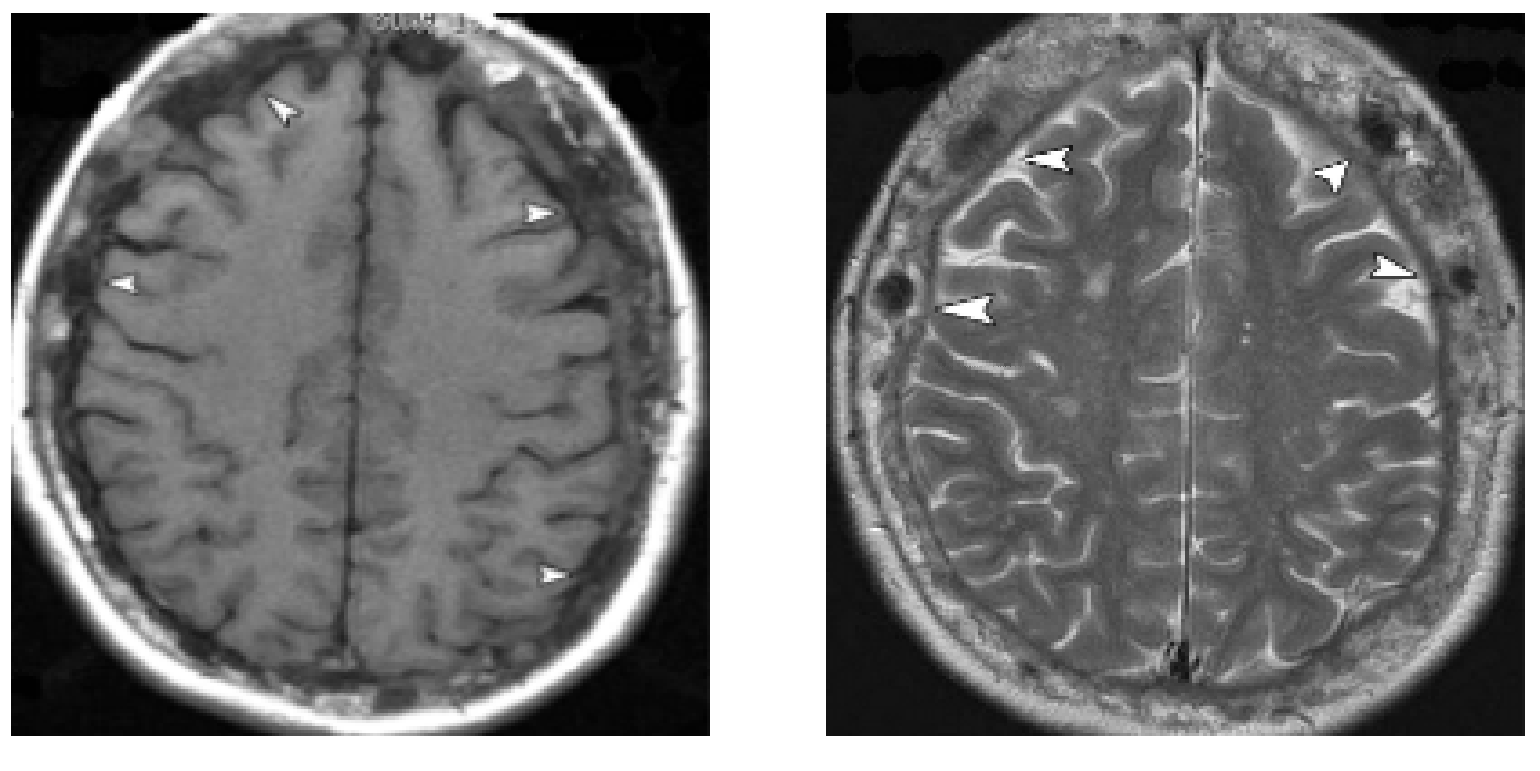
The MRI signal intensity characteristics in Paget disease are variable, reflecting the natural course of the disease process in different phases. Three major patterns of involvement are recognized:
- The most common pattern is dominant signal intensity in pagetic bone similar to that of fat; this pattern of involvement presumably corresponds to long-standing disease and is noted in most patients.
- The second most common pattern corresponds to the early mixed active phase when involved bone shows heterogeneous, relatively low T1 signal intensity and high T2 signal intensity. Referred to as the “speckled” appearance
- The least common pattern of signal intensity changes is seen in the late blastic inactive phase when pagetic bone shows low signal intensity on both T1- and T2-weighted images, suggesting the presence of compact bone or fibrous tissue
MANAGEMENT
- Calcitonin - inhibits bone resorption and provides timely pain relief.
- The current mainstay of treatment in Paget disease -is the second-generation bisphosphonates (i.e., disodium pamidronate, alendronate, risedronate), which are potent inhibitors of bone resorption
- The greatest advantage of second-generation bisphosphonates over calcitonin is prolonged remission of the disease in addition to a more dramatic decrease in the parameters of bone turnover compared with calcitonin.
- Mithramycin, a cytotoxic antibiotic - is best reserved for those cases resistant to other forms of medical treatment.
- Surgical treatment, total joint replacement, especially total hip and knee replacement, has resulted in marked relief of pain and improved locomotion
REFERENCES
- The spine in Paget’s disease
- C Dell’Atti et al
- DOI-Link: https://pmc.ncbi.nlm.nih.gov/articles/PMC1934928/
- Imaging of Paget Disease of Bone and Its Musculoskeletal
- Complications: Review
- Daphne J. Theodorou, et al https://doi.org/10.2214/AJR.10.7222
Dr. RAHUL KARTHIK LINGUTLA
Consultant Radiologist
Manipal Hospital, Yeshwanthpur, Bengaluru.
Dr. NIKITHA U N
Radiology resident
Manipal Hospital, Yeshwanthpur, Bengaluru.