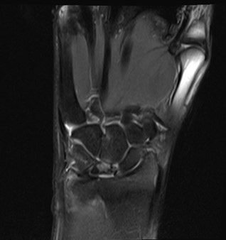
22 year male, History of fall long back, now presents with right wrist joint pain
FINDINGS –
- A. MR RIGHT WRIST
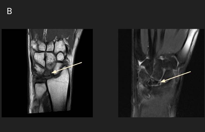
- B. MR RIGHT WRIST
LEGENDS -
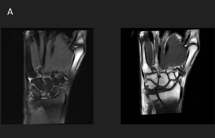
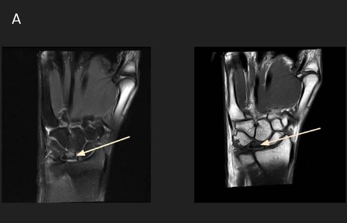
- A. MRI RIGHT WRIST-Volume loss of lunate bone (predominantly along the radial aspect) is seen with altered marrow signal intensity appearing heterogeneously T1 and STIR hypointense. Mild STIR hyperintensity is seen along dorsal aspect of the bone. There is collapse of lunate in the coronal plain
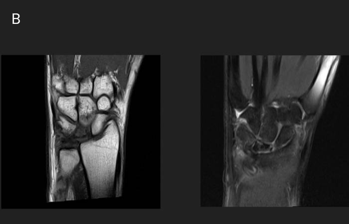
- B. MR RIGHT WRIST-Chronic degenerative partial tear of scapholunate ligament is seen, with mild widening of scapholunate interval.
DIAGNOSIS-
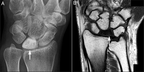
KIENBOCK’S DISEASE
INTRODUCTION-
- Avascular necrosis of the lunate bone leading to progressive collapse and wrist arthritis.
- Most common in young adult males (20–40 yrs)
- Usually dominant wrist
ETIOLOGY AND RISK FACTORS
Proposed Causes
- Repetitive trauma to wrist
- Vascular insufficiency of lunate
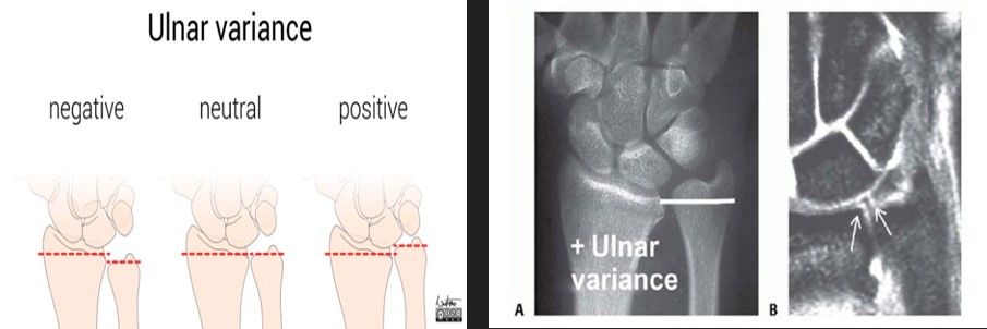
- Negative ulnar variance
- Mechanical overload
Predisposing Factors
- Single palmar vascular supply to lunate
- Carpal instability
- Occupational repetitive loading
ULNAR VARIANCE
KIENBOCK'S VS ULNAR IMPACTION
| Feature | Ulnar Impaction Syndrome | Kienbock Disease |
|---|---|---|
| Ulnar variance | Positive | Negative |
| Pathology | Degenerative overload | Avascular necrosis |
| Bones involved | Lunate + Triquetrum | Only Lunate |
| TFCC | Degeneration / tear common | Usually normal |
| X-ray findings | Sclerosis in lunate & triquetrum | Lunate sclerosis, collapse |
| MRI edema | Lunate + Triquetrum | Lunate only |
| Lunate collapse | Rare early | Common later |
| Carpal instability | Uncommon | Common in advanced stage |