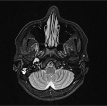
A 37-year-old male presents with progressive right sided hearing loss and a persistent sense of ear fullness for several months.
HISTORY :
- A 37-year-old male presents with progressive right sided hearing loss and a persistent sense of ear fullness for several months.
- MRI temporal bone advised.
FINDINGS :
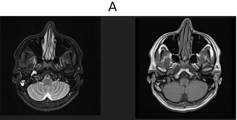
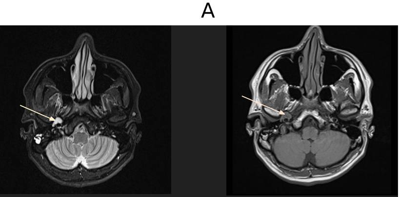
- A. MR TEMPORAL BONE
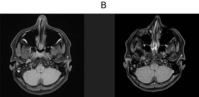
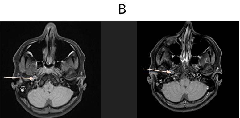
- B. MR TEMPORAL BONE
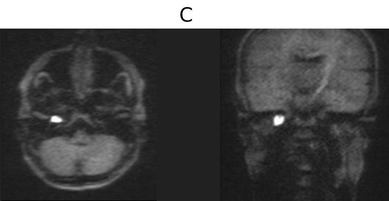
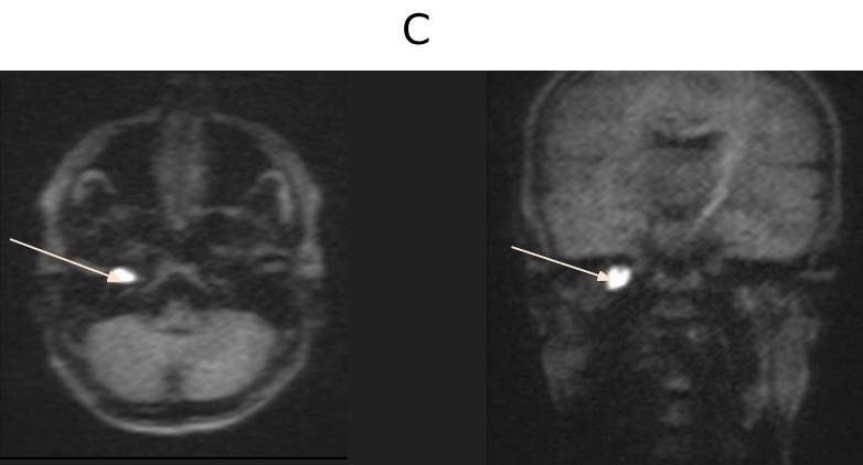
- C. MR TEMPORAL BONE
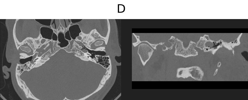
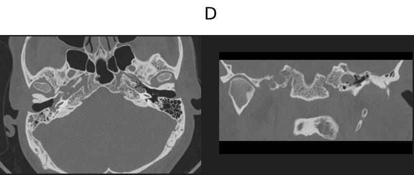
- D. CT TEMPORAL BONE
LEGENDS :
- A. MR TEMPORAL BONE
- A well circumscribed lobulated lesion demonstrating T2/STIR hyperintensity, T1 isointensity measuring ~8 x 12 x 11 mm (AP X TR X CC) in the anteroinferior aspect of the right petrous.
- The lesion lies in the bony portion of the eustachian tube, medial to the temporomandibular joint,lateral to the carotid canal and just posterior to the foramen spinosum.
- B. MR TEMPORAL BONE
- There is no post contrast enhancement.
- C. MR TEMPORAL BONE
- The lesion demonstrates marked restricted diffusion.
- D. CT TEMPORAL BONE
- There is scalloping of bony part of eustachian tube with no erosisons.
DIAGNOSIS
A well circumscribed lobulated lesion in the bony portion of the RIGHT eustachian tube causing marked scalloping of the carotid canal with marked restricted diffusion and no enhancement.
The differential for this include cholesteatoma and an epidermoid cyst.
CHOLESTEATOMA
Definition: Keratinizing squamous epithelium cystic lesion within temporal bone
Types
- Congenital – embryonic epithelial rests
- Acquired – retraction pocket / chronic otitis media
Common Sites
- Middle ear
- Mastoid
- Protympanum
Radiological Features
CT: Soft tissue mass with bone erosion/scalloping
MRI:
- T1: Hypo–iso
- T2: Hyperintense
- DWI: Marked restriction (key feature)
- Post-contrast: No enhancement
Complications
- Ossicular destruction
- Facial nerve canal erosion
- Carotid canal involvement
- Intracranial extension
EPIDERMOID CYST Definition
- Congenital ectodermal inclusion cyst containing keratin debris
Common Sites
- Cerebellopontine angle (most common)
- Parasellar / suprasellar cisterns
Radiological Features
- CT: CSF-like density lesion
- MRI:
- T1: Hypointense (CSF-like)
- T2: Hyperintense (CSF-like)
- FLAIR: Incomplete suppression (“dirty CSF”)
- DWI: Marked restriction (key feature)
- Post-contrast: No enhancement
Growth Pattern
- Insinuating / encasing adjacent vessels and nerves
- Non-expansile compared to cholesteatoma
Complications
- Cranial nerve compression
Management
- Surgical excision (often subtotal due to adherence)
IMAGING FEATURES
| IMAGING FEATURES | CHOLESTEATOMA | EPIDERMOID CYST |
|---|---|---|
| Location | Middle ear, mastoid, Eustachian tube | CPA cisterns, parasellar |
| Margins | Well-defined, lobulated | Irregular, insinuating |
| Bone (CT) | Erosion / scalloping common | Minimal remodeling |
| CT Density | Soft tissue | CSF-like |
| T1 MRI | Hypo–iso | Hypo (CSF-like) |
| T2 MRI | Hyperintense | Hyperintense (CSF-like) |
| DWI | Marked restriction | Marked restriction |
| ADC | Low | Low |
| FLAIR | Variable | Incomplete suppression (dirty CSF) |
| Post-contrast | None | None |
| Growth pattern | Expansile | Encasing / infiltrative |
REFERENCES
CASE CONTRIBUTORS
DR. ANITA NAGADI
SENIOR CONSULTANT RADIOLOGIST,
MANIPAL HOSPITAL, YESHWANTHPUR
DR. FATHIMATH ASHILI KM
RADIOLOGY RESIDENT,
MANIPAL HOSPITAL, YESHWANTHPUR