27 year old lady presents with complains of a slow growing swelling over the wrist which is painless.
HISTORY AND CLINICAL PRESENTATION
- 27 year old lady presents with complains of a slow growing swelling over the wrist which is painless.
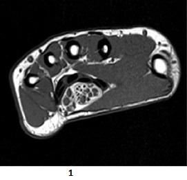
AXIAL T1

Low intensity tubular structures representing nerve fascicles with interspersed high signal representing fat
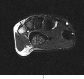
AXIAL T2

Intermediate signal longitudinaly oriented structures representingnerve fascicles
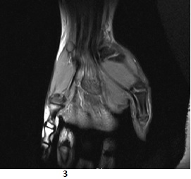
PDFS COR

Fusiform dilatation of median nerve with longitudinally arranged structures representing nerve fascicles giving sphagetti like appearance.
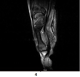
PDFS COR

Fusiform dilatation of median nerve
DIAGNOSIS:
Axial T1 weighted images show bulky median nerve with low signal intensity tubular structures representing thickened nerve fascicles with interspersed high signal representing fat. This appearance is known as Coaxial cable like appearance which is pathognomonic for fibrolipomatous hamartoma of median nerve.
DISCUSSION:
- Fibrolipomatous hamartomas are benign tumours usually affecting infants and less commonly children and young adults.This uncommon lesion is also known as neural fibrolipoma ,lipofibromatous hamartoma ,perineural lipoma and intraneural lipoma.1,2
- The median nerve is overwhelmingly the most common affected nerve (80% of cases), followed by radial,ulnar and dorsum of foot and brachial plexus.3
- Radiographic features-
- Ultrasound- A characteristic hypoechoeic coaxial cabling encased by an echogenic substratum may be seen .
- MRI – MRI features are pathognomonic and typically shows a coaxial cable like appearance on axial images and a spaghetti like appearance on coronal images.
- T1- the neural bundles were hyperintense to muscle and the surrounding substratum was isointense to muscle.
- T2- fat components are high signal and fibrous components are low signal.
REFERENCES
- Cavallaro MC, Taylor JAM, Gorman JD, Resnick D. Imaging findings in a patient with fibro-lipomatous hamartoma of the median nerve.AJR.1993;161:837-838
- De Maeseneer M,Jaovisidha S, Lenchik L.Fibrolipomatous hamartoma:MR imaging findings – Skeletal Radiol.1997;26:155-160
- Murphey MD, Smith WS, Smith SE, Temple HT.From the archives of the AFIP: Imaging of musculoskeletal neurogenic tumors –Radiologic-Pathologic Correlation.Radiographic.1999;19:1253-1280
DR.SHASHWAT PRIYADARSHI
Cross-sectional imaging fellow MHRG